Abstract
The skin is a touch-sensing organ that grows and thickens from birth to adulthood. The touch-sensing cells of the nervous system interact with skin cells and must remodel to accommodate skin growth. In adult animals, the skin also develops specialized structures, like scales in fish and hair in mammals. We studied how changes in the skin and touch-sensing nervous system are controlled, using fish as a model. We found that, in fish mutants with no scales, the outer skin fails to mature and the adult touch-sensing nervous system does not fully develop. As a result, fish without scales are likely less sensitive to touch. These results show that scales instruct cells in the skin and nervous system to transform into adult forms. Our findings in fish raise the possibility that other structures, like hair in mammals, play similar roles in maturing the skin of other animals.
The Skin and Nervous System Carry Out Touch Sensation
Our sense of touch allows us to enjoy pleasurable textures, like a soft pillow, and avoid dangers, like sharp objects or hot substances. Two types of cells in our bodies function together to carry out touch sensation: (1) the cells of the outer skin, called epidermal cells, and (2) specific kinds of nervous system cells, called touch-sensing neurons (Figure 1). Epidermal cells pack tightly together like tiles, to form thin sheets. Several layers of these sheets form a tissue called the epidermis, which covers the surface of our bodies. Touch-sensing neurons are located deep inside our bodies, but send out two long extensions, called axons, to communicate with distant tissues. One of these axons extends to the brain, the other to the skin. In the skin, axon endings squeeze between epidermal cells and branch to form a structure resembling an antenna. This part of the axon also functions like an antenna: it is activated when the skin is touched. Activated axons send electrical impulses to the brain, making animals aware that they touched an object. As an animal grows, maintaining the interactions between epidermal cells and touch-sensing neurons is critical to the animal’s ability to sense touch.
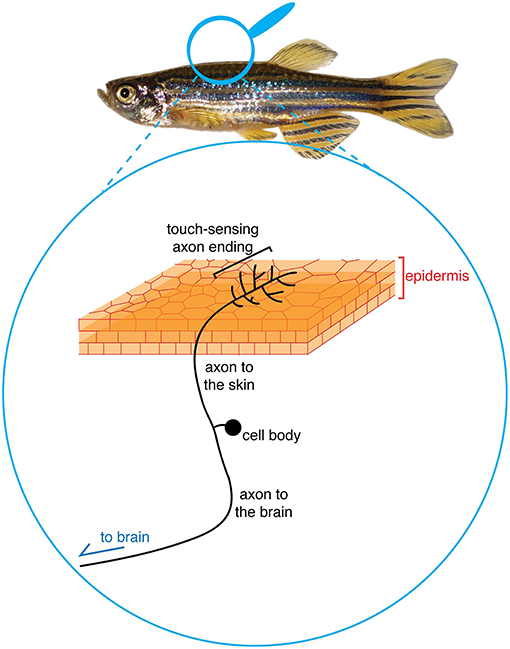
- Figure 1 - Diagram of the cells required for touch sensation.
- Epidermal cells pack tightly together into layers that form the epidermis. The epidermis wraps the outside of an animal’s body. Touch-sensing neurons have two axons: one that goes to the epidermis and another that goes to the brain. Touch is sensed by the branched endings of axons in the epidermis, which send a signal that travels down the axons to the brain.
The Skin Changes From Birth to Adulthood
Animals can sense touch as soon as they are born, even though the skin of a newborn is very simple. The epidermis of a newborn consists of only one or two sheets of epidermal cells, and a small number of touch-sensing neurons send axons to the epidermis. As newborn animals mature into adults, the skin grows to cover the much larger animal, the epidermis thickens by adding more layers of cells, and new touch-sensing neurons are born. In addition to growth and thickening, during this time special structures—like hair in mammals, feathers in birds, or scales in fish—form within the skin. We refer to this maturation as a “metamorphosis,” a process between birth and adulthood when multiple tissues change at once. For example, the transformation of tadpoles into frogs is a metamorphosis. People undergo a similar metamorphosis as teenagers: as we develop from children into adults, many of our tissues grow and change. Our study aimed to understand how epidermal cells and touch-sensing neurons change as the skin undergoes metamorphosis [1].
Fish are an Excellent Model to Study How the Skin Changes as Animals Grow
We chose to use zebrafish, a small tropical fish, for our experiments, since they are easy to study and grow to adulthood in just 3 months. Zebrafish are a popular “model” animal for biological studies (along with mice, fruit flies, and worms), because zebrafish cells and tissues are similar to human cells and tissues, and scientists have developed many tools to study them. Similar to the way many mammals, such as mice, are born without hair, zebrafish are born without scales (Figure 2). Scales are bony discs that form just under the epidermis [2]. Scales are arranged in a regular, overlapping pattern, like roof tiles, along a fish’s body. The epidermis is draped over scales and covered by a protective slime. When fishermen remove a fish’s hard scales to cook it, they are also removing the thin epidermis on top of scales. Scales begin forming when zebrafish are about 1 month old. We think of fish at this age as “teenagers,” since their tissues are undergoing a transition from childhood to adulthood.
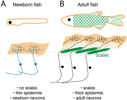
- Figure 2 - The skin and touch-sensing nervous system change as newborn animals grow into adults.
- (A) In newborn fish, the epidermis is thin and contains axon endings of touch-sensing neurons. (B) In adult fish that have undergone metamorphosis, the epidermis is thicker and a different population of touch-sensing neurons now extends axons into the epidermis.
To examine epidermal and touch-sensing cells in detail, we used transgenic zebrafish. Transgenic animals are engineered in a lab to have extra genes in their DNA. Our transgenic fish had genes added to their DNA that instruct epidermal cells or touch-sensing neurons to make a green fluorescent protein (GFP) from jellyfish. When a specific kind of light is shined on cells with GFP, those cells glow green under a microscope. Examining these transgenic animals under a high-powered microscope allows us to see the detailed structure of epidermal cells and neurons.
In zebrafish, major changes in the epidermis and touch-sensing neurons occur around the time that scales form. When fish are born, the epidermis consists of just two epidermal layers, but in adults, the epidermis contains many more layers (Figure 2). Newborn and adult fish have different populations of touch-sensing neurons–the touch-sensing neurons in newborn fish eventually die and are replaced by a different set of adult touch-sensing neurons. Since these changes in the epidermis and touch-sensing neurons happen when scales are forming (when fish are “teenagers”), we hypothesized that scales help to cause changes in the epidermis and sensory neurons, as animals grow to adulthood.
Experiment 1: Do Scales Promote Changes in the Skin?
To test our hypothesis, we examined the skin of mutant fish with no scales [3]. These fish are mutants because they have a defect in a gene required to form scales. Without scales, fish would probably not survive in the wild, but in the controlled environment of our laboratory they survive into adulthood. We created fish that were both transgenic (they had genes for making GFP in epidermal cells or neurons) and mutant (they lacked genes needed for making scales). When we examined epidermal cells and neurons in these mutant fish, we discovered that they had several defects (Figure 3). First, in adult mutant fish without scales, the epidermis was thinner than in normal adult fish. Second, the branched endings of touch-sensing neurons were missing from the epidermis of mutants. In newborn mutant fish, touch-sensing neurons were normal, and these neurons were replaced by adult neurons, even though scales never formed. However, these adult touch-sensing neurons failed to extend their antenna-like axon endings into the epidermis of the mutants. Without axons in the epidermis, these fish probably cannot sense touch normally. This experiment showed that our hypothesis was correct—scales are required for changes to occur in the epidermis and touch-sensing neurons of “teenage” fish.
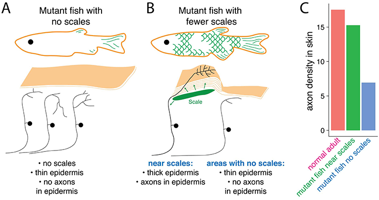
- Figure 3 - Scales send a local signal to transform epidermal cells and touch-sensing neurons.
- (A) In mutant fish that do not have scales, the epidermis stays thin and the axons of touch-sensing neurons do not enter the skin. (B) In mutants missing only some scales, the epidermis matures only in areas near scales. In areas without scales, the epidermis remains thin and touch-sensing axons do not enter it. The arrows represent signals that scales send to surrounding cells. (C) Graph showing the axon density in the skin of normal adults, and in areas with and without scales in mutant adults. The Y-axis shows arbitrary units representing relative axon density. This graph shows that areas of the epidermis near scales have more axons than areas far from scales.
Experiment 2: Do Scales Send Short- or Long-Range Signals to the Epidermis and Neurons?
One explanation for our findings is that scales send signals to the epidermis and touch-sensing neurons, telling them to change into adult forms. These scale signals instruct epidermal cells to multiply and form more layers. They also instruct adult touch-sensing neurons to send axon endings into the epidermis. Without scales, these signals are missing, so the epidermis and touch-sensing neurons fail to change like they would in normal fish.
We wondered if scale signals act at long or short distances. If they act at a long distance, just a few scales might be able to promote changes to the epidermis and touch-sensing neurons across the entire animal. By contrast, if scale signals act at a short distance, they would only reach nearby epidermal cells and neurons, causing only those regions to have a thicker epidermis with touch-sensing axon endings. To distinguish between these possibilities, we examined another set of fish mutants. These mutant fish lacked just a few scales, rather than all of their scales (Figure 3) [4]. In these fish, we compared regions of the body with scales to regions of the body without scales. In these mutants, the epidermis in areas with scales was thick, like in normal adults, and full of axon endings. However, in areas without scales, the epidermis was thin and lacked axon endings. We concluded that scales normally send a short-range signal to thicken the epidermis and attract axons, just to areas close to the scale. We do not know what this signal is, but a major goal of our future research is to identify substances on scales that transform the epidermis into its adult form.
Conclusions and Implications: Do Our Results in Fish Apply in Other Animals?
Our experiments showed that scales are critical for changing the skin from its newborn form to its adult form. Scales send a signal to nearby epidermal cells, telling them to grow and thicken the epidermis, and a signal to adult touch-sensing neurons, telling them to send axons into the epidermis. By promoting changes in both the epidermis and axons, scales ensure that these two metamorphic events happen together, allowing fish to develop adult skin that carries out normal touch sensation.
Do our results apply only to fish, or might they provide a clue to similar processes in mammals, including humans? Like in fish, as we develop from fetuses into adults, our skin thickens and axons of our touch-sensing neurons enter the epidermis. How these processes occur in humans is not understood. Humans obviously do not have scales, but most of our skin contains hair. Similarly, the skin of birds contains feathers. Hairs, feathers, and scales are very different structures, but early in their formation they share similarities, revealing that they are related to one another in evolution. Our results suggest that hairs and feathers may function like scales to promote skin maturation, and to help distribute the endings of touch-sensing neurons into the epidermis. We hope our study inspires other scientists to test if hairs or feathers promote such changes in mammals or birds, like scales do in fish.
Glossary
Neurons: ↑ The major cells of the nervous system, also called nerve cells. Neurons communicate with each other to allow us to sense the environment, move, learn, and think.
Epidermis: ↑ The outermost part of the skin, made up of epidermal cells tightly packed together into sheets and layers.
Axons: ↑ Thin, wire-like projections from neurons that often extend long distances. Axons conduct electrical impulses to communicate with distant cells.
Metamorphosis: ↑ A major transition in animal development. During metamorphosis, multiple tissues remodel at once. The transformation of caterpillars into butterflies, or tadpoles into frogs, are dramatic examples of metamorphosis.
Transgenic Animals: ↑ Animals engineered to have extra genes in their DNA. We used transgenic fish expressing a fluorescent protein gene from jellyfish in specific cells, making detailed cellular structures visible under a microscope.
Conflict of Interest Statement
The authors declare that the research was conducted in the absence of any commercial or financial relationships that could be construed as a potential conflict of interest.
Original Source Article
↑Rasmussen, J. P., Vo, N.-T., and Sagasti, A. 2018. Fish scales dictate the pattern of adult skin innervation and vascularization. Dev. Cell. 46:344–59.e4. doi: 10.1016/j.devcel.2018.06.019
References
[1] ↑ Rasmussen, J. P., Vo, N.-T., and Sagasti, A. 2018. Fish scales dictate the pattern of adult skin innervation and vascularization. Dev. Cell. 46:344–59.e4. doi: 10.1016/j.devcel.2018.06.019
[2] ↑ Sire, J.-Y., and Akimenko, M.-A. 2004. Scale development in fish: a review, with description of sonic hedgehog (shh) expression in the zebrafish (Danio rerio). Int. J. Dev. Biol. 48:233–47. doi: 10.1387/ijdb.031767js
[3] ↑ Harris, M. P., Rohner, N., Schwarz, H., Perathoner, S., Konstantinidis, P., and Nüsslein-Volhard, C. 2008. Zebrafish eda and edar mutants reveal conserved and ancestral roles of ectodysplasin signaling in vertebrates. PLoS Genet. 4:e1000206. doi: 10.1371/journal.pgen.1000206
[4] ↑ Rohner, N., Bercsényi, M., Orbán, L., Kolanczyk, M. E., Linke, D., Brand, M., et al. 2009. Duplication of fgfr1 permits Fgf signaling to serve as a target for selection during domestication. Curr. Biol. 19:1642–7. doi: 10.1016/j.cub.2009.07.065