Abstract
The brain controls the activities of the body, including food digestion, drinking, sleep cycles, temperature, blood pressure, and more. These functions are essential to keep the body in homeostasis, which is the state of being steady and balanced. To control homeostasis, the brain talks to the body with the help of chemical messengers called hormones. Hormones travel through the blood stream from the brain to the body and back. However, in order to protect the delicate brain cells from unwanted intrusions, the blood vessels of the brain are tightly sealed, preventing the passage of most molecules. How, then, does the brain bypass this barrier to communicate with the body? The answer is that, in certain parts of the brain, the blood vessels contain special window-like openings that allow passage of hormones. Scientists are investigating why and how some blood vessels open their windows while others remain sealed.
Keeping the Body Balanced and Steady
The outside world in which we live is constantly changing. The inside of our bodies also changes after we eat, drink, exercise, or sleep. Yet, despite these continuous changes, the body is able to keep its inner environment stable. We call this ability homeostasis, which is a combination of two words in Greek: “homeo,” meaning “similar,” and “stasis” meaning “stable.” For example, all the cells in the human body function best at a temperature of around 37°C. Therefore, the body works to maintain this temperature. When it is hot outside, we cool the body by sweating. When it is cold outside, we warm up by shivering, which produces heat. If we were unable to control body temperature, our cells would fail to function properly. That is why all living organisms, from single-celled bacteria or yeast to large animals, such as elephants, must maintain homeostasis to stay alive.
Homeostasis is achieved by a never-ending “conversation” that takes place between the brain and the rest of the body. The brain, which serves as the main headquarter of the body, receives information about the state of the many organs and tissues. Then it “decides” what needs to be done in response to this information (Figure 1). If homeostasis is disrupted, the brain sends commands to the various body parts to bring bodily conditions back to normal. The language of this conversation is chemical. Special molecules called hormones serve as messengers that carry information and instructions back-and-forth between the brain and the body, as well as between various body parts. The circulatory system, which carries the blood, is the main route of communication. This elaborate network of pipe-like structures called blood vessels runs through the entire body, including the brain. The passage of hormones and other signaling molecules between the blood and the body tissues occurs at very small vessels called capillaries.
- Figure 1 - The brain maintains the body’s well-being.
- A constant conversation between the brain and the rest of the body takes place to keep physical conditions steady and balanced—a state called homeostasis.
For example, let us look at what happens after we have a meal. After we eat, the amount of a sugar called glucose is increased in the blood. An organ called the pancreas senses the elevated level of glucose and sends a hormone message to the brain. You may have heard of this hormone; it is called insulin. After receiving the message from the pancreas, the brain guides the liver to absorb the excessive sugar from the bloodstream. The end result is that blood sugar levels get back to normal. At the same time, the gut sends other hormones to the brain to announce that the belly is full. The brain reacts by sending a hormone that delivers a “stop eating” signal.
The Hypothalamus: the Brain’s Center of Homeostasis
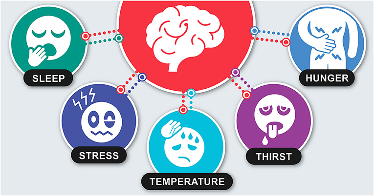
So far, we have described how the brain collects information from the body and decides which commands to send in order to maintain homeostasis. The specific region of the brain where most of this activity takes place is called the hypothalamus, which means “under the inner chamber” in Greek (Figure 2). The hypothalamus controls many important body functions, such as sleep, blood pressure, temperature, hunger, thirst, and energy consumption and storage. Much like a computer microprocessor, the hypothalamus runs an algorithm that computes the information by following a set of rules. Then, the hypothalamus makes a decision about whether or not to send commands to the body. This type of computation occurs in brain cells called neurons. The neurons in the hypothalamus can receive feedback both from inside the body and from the external environment. They can also produce various hormones.
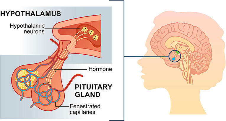
- Figure 2 - The meeting point between hypothalamic neurons and pituitary capillaries.
- On the right, the hypothalamus is highlighted in green and the pituitary in blue. On the left, you can see that commands from the hypothalamus travel along the neurons to the pituitary gland. The pituitary then releases commands in the form of hormones into the bloodstream, via the fenestrated capillaries.
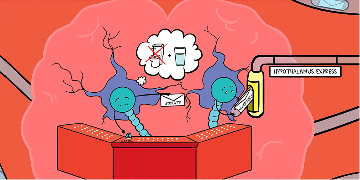
As an example from our daily lives, when we do not drink enough water on a hot day, the body sends a message to the brain which, in turn, releases a hormone called vasopressin into the blood. Vasopressin instructs the kidneys to soak water back up into the blood. This prevents us from urinating and protects us from further loss of water. Simultaneously, hormones from the brain make us feel thirsty so that we drink more and replenish our water supplies.
Windows Between the Brain and Blood
As mentioned, the bloodstream acts like a postman, collecting and delivering chemical messages to and from the brain. However, there is a major obstacle in the postman’s path. The blood vessels of the brain are sealed tightly by a special safeguard, called the blood-brain barrier (BBB). The cells that build the blood vessel walls are connected tightly to one another, similar to bricks glued together by cement, so almost nothing can pass through (Figure 3, bottom). The main purpose of this barrier is to protect the brain from infection. When the BBB is loosened as a result of disease or injury, bacteria might enter the brain and cause severe damage or even death.
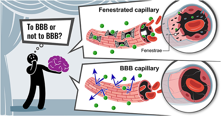
- Figure 3 - To BBB or not to BBB?
- There are two types of capillaries in the brain. On the top, you can see fenestrated (windowed) capillaries lacking the BBB. The wide-open windows in the capillary wall allow passage of molecules (green) to and from the bloodstream. On the bottom, you can see a tightly sealed BBB capillary, which restricts entry of molecules to keep brain cells safe. v
Protecting the brain is very important, but if the brain’s blood vessels are firmly sealed by the BBB, how does the hypothalamus communicate with the body to maintain homeostasis? The answer is that there are unique brain regions that lack the BBB and, instead, have special leaky blood capillaries. One of such leaky regions is an organ called the pituitary, which is located at the bottom of the brain [1] (Figure 2). Looking at the pituitary’s capillaries under a microscope with an extremely high magnification reveals that they have very small openings called fenestrae [2], which is the Latin word for windows (Figure 3, top). These tiny windows are ten thousand times smaller than the tip of a hair strand! Fenestrae allow the rapid passage of molecules, such as hormones, between the brain and the blood circulation. Thus, commands that are generated in the hypothalamus travel to the pituitary and meet the fenestrated blood vessels (Figure 2). There, the commands are released to the bloodstream to reach the organs.
A Dilemma: to BBB or not to BBB?
Areas of the brain that contain fenestrated blood vessels have been known to scientists for quite a while. However, it is still unclear how the vessels in these regions remain leaky. The blood vessels of the brain face a dilemma: to make fenestrae that enable communication with the body at the risk of infection, or to protect the brain from harmful invaders by isolating it. We sometime call this dilemma “To BBB or not to BBB?” after the famous quote from William Shakespeare’s play Hamlet: “To be or not to be, that is the question” (Figure 3).
We recently investigated the unique fenestrated vessels located where the neurons of the hypothalamus make contact with the pituitary (Figure 2). We found that blood vessels in this region receive cues from neighboring cells called pituicytes, telling them to form fenestrae [3, 4]. Pituicytes have been known for many years to help with the release of commands from the neurons of the hypothalamus into the bloodstream. We identified two types of signaling molecules produced by the pituicytes that make the blood vessels in this part of the brain leaky. One type tells the blood vessels to form the fenestrae and thereby remain leaky, while the other type blocks the formation of the BBB. When these signaling molecules are blocked, the blood vessels in the pituitary stop making fenestrae and instead start forming the BBB. This means that the pituicytes ensure that the windows are open using two forms of control. This is a bit like wearing both a belt and suspenders to hold your jeans in place. This double control guarantees the free passage of hormones into the bloodstream.
Why is This Important?
Fenestrated blood vessels serve as important gateways that allow communication between the brain and the blood. However, blood vessels with fenestrae are also found in other important organs, such as the pancreas, liver, and kidneys. In these organs, it is also necessary to exchange molecules with the blood circulation. Yet we still know very little about how fenestrae are formed. Revealing the secrets of window-making could be very useful. For example, while the BBB safeguards the brain from infection, it also prevents the passage of medicines into the brain. This makes it difficult for doctors to treat brain diseases. If we find a way to create fenestrae in the tightly sealed blood vessels of the brain, it may be possible to deliver drugs across the BBB to treat disorders, such as epilepsy, Parkinson’s disease, and autism.
Glossary
Homeostasis: ↑ A process by which an organism maintains inner environment stable while adjusting to conditions that are best for its survival.
Hormones: ↑ Molecules produced by glands and the brain, which serve as chemical messengers. Hormones are transported by the blood to organs all over the body, to regulate the body’s function.
Capillary: ↑ The smallest type of blood vessel, which delivers nutrients and oxygen to all cells of the body.
Neuron: ↑ A basic unit of the nervous system, also called a nerve cell. Neurons can process and transmit information to other nerve cells and to other cell types, such as muscle cells.
Blood-brain Barrier: ↑ Physical barrier within the blood vessels of the brain that prevents substances from passing into the brain, keeping the environment of the brain safe and stable.
Fenestrae: ↑ Tiny openings in capillary walls that allow passage of molecules to and from the bloodstream. Fenestrated capillaries are found in organs where the rapid exchange of information between the blood and the tissue is required (pancreas, intestines, some brain regions, etc.).
Pituicytes: ↑ Specialized cells of the pituitary gland that help the release of hormones from neurons into the bloodstream. Pituicytes have recently been shown to help keep pituitary blood vessels leaky.
Conflict of Interest
The authors declare that the research was conducted in the absence of any commercial or financial relationships that could be construed as a potential conflict of interest.
Acknowledgments
We thank Genia Brodsky and Keren Katzav for their valuable help in illustrations preparation. We thank Nitzan Konstantin for English editing.
References
[1] ↑ Gutnick, A., and Levkowitz, G. 2012. The neurohypophysis: fishing for new insights. J. Neuroendocrinol. 24:973–4. doi: 10.1111/j.1365-2826.2012.02292.x
[2] ↑ Gordon, L., Blechman, J., Shimoni, E., Gur, D., Anand-Apte, B., and Levkowitz, G. 2019. The fenestrae-associated protein Plvap regulates the rate of blood-borne protein passage into the hypophysis. Development 146:dev177790. doi: 10.1242/dev.177790
[3] ↑ Anbalagan, S., Gordon, L., Blechman, J., Matsuoka, R. L., Rajamannar, P., Wircer, E., et al. 2018. Pituicyte cues regulate the development of permeable neuro-vascular interfaces. Dev. Cell 47:711–26.e5. doi: 10.1016/j.devcel.2018.10.017
[4] ↑ Chen, Q., Leshkowitz, D., Blechman, J., and Levkowitz, G. 2020. Single-cell molecular and cellular architecture of the mouse neurohypophysis. eNeuro 7:ENEURO.0345-19.2019. doi: 10.1523/ENEURO.0345-19.2019