Abstract
Did you know that it is not only your eyes that allow you to see? Your brain functions with the eyes to process and make sense of all things that you see. A part of the brain called the visual cortex is responsible for vision. The brain contains over 100 billion brain cells called neurons, and they work in “levels” to help you see the world—from a basic level in which you perceive simple shapes up to higher levels where you understand complex patterns. When networks of brain neurons do not work properly, brain disorders can result. Doctors and scientists can use various techniques to measure the activity of neurons. For example, unusual patterns of brain waves can tell us about damaged neural networks and brain abnormalities. Computers can also be programmed to “see” visual information, and such computers can help us to learn about the vision process in humans.
How Do You See the World?
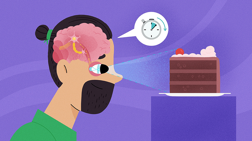
Do you like the world-famous Korean band BTS? In the case of the band, BTS stands Bang Tan Sonyeondan, but in neuroscience, it could also stand for brain transfer stimulus, which is a crucial brain function. The brain recognizes what your eyes see and can interpret those visual information or stimulus, so you can understand what you are reading or seeing. A stimulus is anything that captures your attention. When we are talking about vision, you detect visual stimuli, such as light. For instance, when you are at a BTS concert, you see powerful lights flashing with various colors, but this does not surprise you. The brain receives these visual stimuli and processes them with other visual information, so that you know the lights come from the stage. In other words, your brain helps you to recognize things, such as stage lights and a singer at a concert.
How Does Your Brain Help You See?
Have you ever wondered how your eyes allow you to see? For instance, when you are at a concert, how can you spot a friend in the huge crowd? Even though you see with your eyes, a part of the brain called the visual cortex is also responsible for vision, as it processes any visual information from the eyes. This brain region contains several levels for analyzing information [1]. Let us think of the visual cortex as a fabulous, multi-layered cake. On the bottom, you have the primary visual cortex (V1), which interprets dots or any forms without shapes. Surprisingly, it only takes 70 milliseconds (msec, 0.07 seconds) to move signals from the eyes to V1.
Next, the secondary visual cortex (V2) recognizes more detailed visual representation than V1, such as geometric shapes. You can think of it this way: as you move up in cake layers, the levels of the visual cortex interpret more complex visual information [2]. With higher levels of visual cortex, you can see different colors or movements. It takes longer to transmit signals from the eyes to “higher” levels of the visual cortex. It takes 100 msec (0.1 seconds) to move from the eyes to V2, about 120 msec (0.12 seconds) from the eyes to V3.
In other words, visual information moves from the eyes through the levels of the visual cortex in a very short amount of time (Figure 1). This explains how you can quickly recognize a friend at a concert. Also, as you are reading this article, you can understand the meaning of each word in a sentence, instead of seeing just a jumble of words. Your brain and eyes work as a team to process visual information.
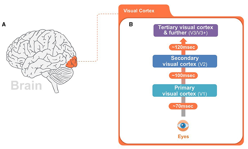
- Figure 1 - Levels of the visual system and the amount of time it takes them to process visual information.
- (A) The visual cortex, the part of the brain that deals with processing the things we see, is shown in red. It is located at the back of the brain. (B) The primary visual cortex, which analyzes very basic visual information like shapes, works the most quickly. The higher levels of visual processing, which recognize more detailed visual information like patterns in V2 and colors and movements in V3/V3+, each take a little longer. Together, visual cortex of the brain receives visual signals from the eyes and tries to make sense of what you are seeing. This is how you can understand a book you are reading or recognize a friend’s face.
How Do We Know How the Human Brain Works?
The human brain has billions of cells called neurons [3]. Neurons connect with each other to form networks, and they communicate using tiny electrical signals. If the networks of neurons in the brain are damaged, neurons cannot receive electrical signals from each other. This can negatively affect brain functions, leading to disorders like memory impairment and dementia.
Since we cannot see the inside of the brain, how do we know if there is an issue with brain networks? Scientists and doctors have several ways to analyze the brain! Since neurons communicate using electrical signals, we can measure the brain’s electrical activity using small, non-metallic devices called electrodes (Figure 2A).
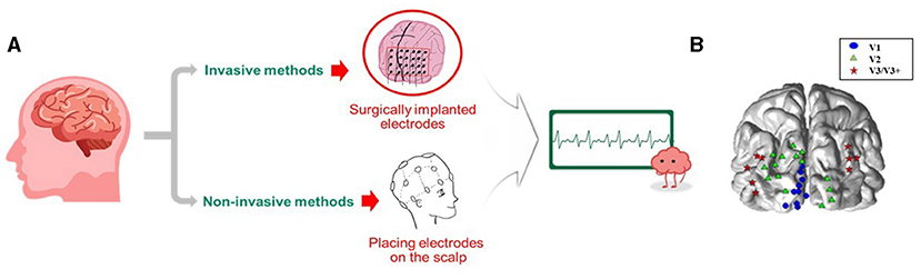
- Figure 2 - (A) To detect electrical signals in the brain, electrodes can either be placed on the scalp or implanted into the brain during surgery.
- Electrodes allow us to measure the brain waves. (B) The brain response to things we see happens in “levels”, with the first level, V1 (blue dots) performs simple visual tasks like recognizing shapes. In an intermediate level, V2 (green triangles) recognizes patterns and colors, and for higher levels, V3/V3+ (red stars) give us complex visual information like colors and movements.
Electrodes help us to see brain waves made from the electrical activities of neurons. Just like waves in the ocean, the brain waves look like wavy lines moving up and down. However, if a person has brain damage, the electrodes might show slow and unusual patterns of brain waves. The ability to accurately read brain waves is important in neuroscience because it helps us to identify brain abnormalities or disorders.
To record the brain activity of patients in our study, we placed electrodes into the visual cortex during surgery, and then showed patients pictures of various shapes and patterns. As patients looked at these visual stimuli, the electrodes recorded their brain waves, so we could examine which brain regions became activated and how the brain responded to each shape and pattern (Figure 2B). For simple visual responses like dots or a flash of lights, V1 regions were activated. For intermediate visual responses, like geometric shapes, such as triangles, circles, V2 areas were activated, and for complex visual responses like visual fantasy or an illusion with mixed colors, V3/V3+ areas were activated.
There are other ways to measure brain activity that do not require surgery, such as electroencephalography (EEG). In EEG, electrodes can be harmlessly placed on the patient’s scalp. EEG is widely used to look at brain activity and identify brain disorders [4].
Today, computers also help us to understand brain abnormalities. Using artificial intelligence (AI), computers can mimic the networks of neurons in the human brain (Figure 3), which allows these computers to function similarly to the brain. For instance, AI-based computerized “vision” allows computers to recognize and interpret visual stimuli, kind of like they are “seeing”. This computer-based “seeing” is similar to human vision, but computers can identify things more quickly and accurately. Thus, whenever we have difficulties in finding brain disorders in patients, we can use AI networks to gain more insight into disorders and treat patients more efficiently [5].
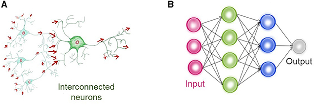
- Figure 3 - (A) Networks of interconnected neurons in the brain work together to help us see.
- Electrical information flows from one neuron to another. (B) AI can simulate the networks of neurons in the human brain, with information flowing between signals (colored circles). In this case, input is a visual stimulus, such as image, and output recognizes what the image is. In fact, artificial neurons in AI look similar but also serve a similar function as neurons in human brain.
Summary: Why Is This Work Important?
In this article, we told you about how the visual cortex functions in vision and how scientists and doctors can monitor brain activities by measuring brain waves. Now you know that the brain must work with the eyes to allow you to see! Networks of brain cells in the visual cortex communicate to process “levels” of visual information, from simple to complex. When brain networks do not work properly, brain disorders like memory impairment and dementia can result. Computerized neural networks, like those used by AI, can help scientists to understand what goes wrong in brain disorders. With our work, we hope to inspire many bright young scientists to show interest in neuroscience and help to answer more fascinating questions about vision someday!
Glossary
Stimuli: ↑ A visual signal that captures an attention.
Visual Cortex: ↑ An area of the brain that processes visual information and has three main layers: primary, secondary, and tertiary.
Neuron: ↑ A nerve cell that communicates with other nerve cells and forms networks to interpret the information that comes in through our senses, like the things we see.
Electrode: ↑ Small non-metallic devices that measure brain electrical patterns are used in both invasive and non-invasive brain imaging techniques.
Brain Waves: ↑ Recordings of electrical signals of the brain.
Electroencephalography: ↑ A non-invasive brain imaging method that measures electrical activities of the brain by placing electrodes on a scalp.
Artificial Intelligence (AI): ↑ A computerized system that mimics the networks of neurons in the brain and can perform human-like tasks.
Neuroscience: ↑ The study of human brain and the interaction of brain cells involved in memory functions and behaviors.
Conflict of Interest
The authors declare that the research was conducted in the absence of any commercial or financial relationships that could be construed as a potential conflict of interest.
Acknowledgments
We confirm that we have read the Journal’s position on issues involved in ethical publication and affirm that this report is consistent with those guidelines. Part of this work was invited to present at Frontiers for Young Minds through Organization for Human Brain Mapping (OHBM) 2021. The paper was supported by the RP-Grant 2021 and 2022 of Ewha Womans University (SEK). The study was supported by grants of the Basic Science Research Program, Convergent Technology R&D Program for Human Augmentation, BK21 Plus Program through the National Research Foundation of Korea (NRF) funded by the Ministry of Science, Information and Communication Technologies & Future Planning [NRF-2018M3C1B8016147, 2019M3C1B8090803, and 2020R1A2C2013216], and Institute of Information & Communications Technology Planning & Evaluation (IITP) funded by the Korea government (MSIT) [No. RS-2022-00155966], and Artificial Intelligence Convergence Innovation Human Resources Development from Ewha Womans University to HL.
Original Source Article
↑Kim, S. E., Kim, W. S., Kim, B. G., Chung, D., Jeong, J., Lee, J. S., et al. 2013 Spatiotemporal dynamics and functional correlates of evoked neural oscillations with different spectral powers in human visual cortex. Clin. Neurophysiol. 124:2248–56. doi: 10.1016/j.clinph.2013.04.341
References
[1] ↑ Gilbert, C. D., Li, W. 2013 Top-down influences on visual processing. Nat. Rev. Neurosci. 14:350–63. doi: 10.1038/nrn3476
[2] ↑ Herculano-Houzel, S. 2009 The human brain in numbers: a linearly scaled-up primate brain. Front. Hum. Neurosci. 3:31. doi: 10.3389/neuro.09.031.2009
[3] ↑ Ghose, G. M., Maunsell, J. 1999 Specialized representations in visual cortex: a role for binding? Neuron. 24:79–85. doi: 10.1016/S0896-6273(00)80823-5
[4] ↑ Kalaivani, M., Kalaivani, V., Devi, V. A. 2014 “Analysis of EEG signal for the detection of brain abnormalities,” in International Journal of Computer Applications® year.
[5] ↑ Hassabis, D., Kumaran, D., Summerfield, C., Botvinick, M. 2017 Neuroscience-inspired artificial intelligence. Neuron. 95:245–58. doi: 10.1016/j.neuron.2017.06.011