Abstract
Pregnancy and childhood are important times of great change and growth. Inside the mom’s body, a baby grows from the size of a poppy seed to the size of a watermelon in just 10 months! After birth, the brain doubles in size by the time the baby is 1 year old. As a child’s body grows, scientists and doctors may need to investigate how things are changing by taking a picture of the inside of the child’s body. With this information, doctors can understand if the child needs special care, and researchers can learn about the growing body. One way to collect such pictures is by using magnetic resonance imaging (MRI). In this article, you will learn how MRI works, when it is helpful to use during pregnancy and childhood, and how researchers around the world are using MRI in new ways.
Magnets are Everywhere!
Magnets have an invisible force that can push or pull certain metal objects. Almost everything that uses electricity or runs with a motor has magnets in it. The magnetic force helps your car run, your microwave cook, and your computer work. Even the Earth we live on is a giant magnet. Magnets are also used to help take pictures of the inside of our bodies to figure out if you are hurt or sick.
How Can Magnets Take Pictures Inside Our Bodies?
Magnetic resonance imaging (MRI) scanners are giant donut-shaped magnets with a tunnel in the center. The magnets are similar to the ones that stick to your fridge or to your white board at school, except the magnets in an MRI scanner are much bigger and stronger. In fact, the magnet in a typical scanner generates a magnetic field 30,000 times stronger than the Earth’s magnetic field! The magnet is so powerful that when you enter the center of the MRI scanner, the water molecules in your body line up with the scanner’s magnetic field. Think of your body’s water molecules as fidget spinners with tiny magnets. The magnetic field of the MRI scanner forces the water molecules to spin in alignment. However, you cannot feel your water molecules moving, and the movement is completely safe for your body.
Next, to create an image, the water molecules must be pushed out of alignment with the magnet’s field. To do this, radio waves are sent through a piece of equipment that acts like an antenna (called an imaging coil). However, rather than making sound waves like those that allow you to listen to music in your car, these radio waves help create signals inside your body. If the radio waves match the frequency of the water molecules (a condition called being “on resonance”, meaning they vibrate together at the same rate), they can exchange energy. When the radio waves are turned off, the water molecules are pulled back to their original positions, releasing energy. The imaging coil measures the change in energy from the water molecules and an image can be created using mathematical calculations.
Each part of your body has different amounts of water molecules giving off different signals. For example, your heart is full of blood and therefore has many more spinning water molecules compared to your knee, which is made of hard bone and thus has many fewer. The differing amounts of energy help to give detail to the image of your body. For more information about how MRI works, we recommend reading this Frontiers for Young Minds article or this one.
Is MRI Safe?
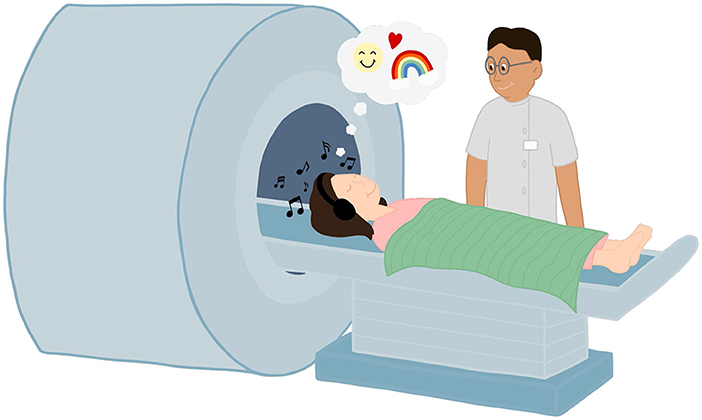
MRI is considered safe for people of all ages—babies, children, adults, and pregnant women. Unlike other imaging procedures such as X-rays or CT scans, MRI does not use any ionizing radiation and, therefore, does not cause any damage or harm to our bodies. However, if you have had any surgery during which a doctor put a magnetic material into your body, the strong magnet in the MRI could make the metal move or cause problems. Usually, plates and screws used during surgery are non-magnetic and will not cause a problem. However, it is still important to tell the MRI technologist collecting the images about any surgeries you have had, so they can decide if they want to use a different test to keep you safe. Also, if you have a pacemaker (a device to regulate your heart), the strong magnet might cause the device to stop working correctly. If you do not like small spaces, the MRI scanner can feel scary and tight because it is a long tunnel. Sometimes scanners have mirrors to give the illusion of more space, to help make people feel more comfortable. Some people choose to close their eyes.
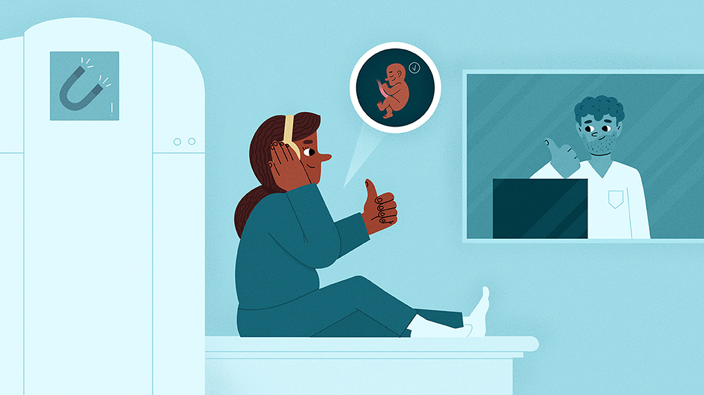
The MRI scanner makes lots of loud noises (as loud as an alarm clock) as it collects the image. You can wear earplugs or headphones to block out the noise. There is also a microphone inside the MRI scanner so you can talk to the MRI technologist. While a parent cannot go into the scanner with you, they can always be nearby. You should talk to your doctor and/or the MRI technologist if you are feeling nervous or scared about having an MRI scan (Figure 1). We, the authors of this article, have had a range of experiences when receiving MRI scans. Some of us said it was painless, relaxing, and even fun. Others said they felt a little uneasy about the small space; however, listening to music helped calm their nerves.
- Figure 1 - The MRI technologist collects the images and will help make you feel comfortable and at ease during the scan.
- You can close your eyes, listen to music, and have a blanket to keep you warm. You have to stay still during the scan, so the picture is not blurry.
MRI During Pregnancy and Childhood
The most typical images collected during pregnancy are from an ultrasound machine, which uses sound waves to create an image of the baby. Unlike an MRI, which requires the patient to go inside a tunnel, an ultrasound scan uses a hand-held probe that is placed on the skin. A gel is used to make the ultrasound images clearer. However, some questions about the health of the baby or the mother are best answered using MRI. Figure 2 shows the differences between an ultrasound and MRI scan of a baby, and Figure 3 provides some examples of how MRI is used during pregnancy.
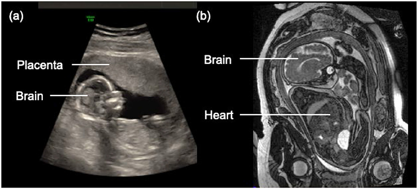
- Figure 2 - (A) Ultrasound image of a baby.
- (B) An MRI image of a baby. The MRI image provides more details about the internal organs (e.g., brain, heart) (image provided by John Sled and Mike Seed at The Hospital for Sick Children).
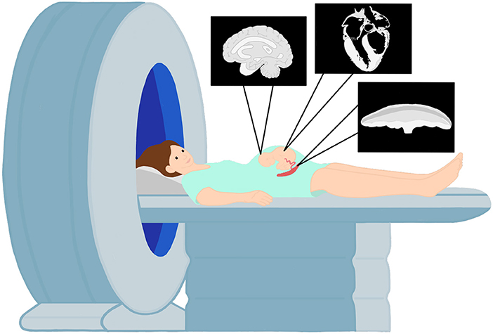
- Figure 3
- MRI is regularly used during pregnancy to provide detailed images of the placenta (which is connected to the baby by the umbilical cord and delivers oxygen and food) and the baby’s brain and heart.
MRI provides excellent images of the baby’s brain and can reveal if there are areas of concern, such as problems with how brain tissue is developing. It can also show if there are issues in the baby’s heart, like a small hole. MRI is also used to study the placenta, the organ that connects the baby to the mother and supplies oxygen and food to the baby. MRI can provide information on whether the placenta is working properly (that is, delivering enough oxygen and food so the baby can grow) and if the placenta is growing in the correct place. A doctor can use MR images collected during pregnancy to make decisions about treatment plans and whether it is time to deliver the baby.
After birth, children might need an MRI scan for many reasons—for example, after an injury, or to help figure out what is wrong if a child is not feeling well. If a child has a hard hit to the head, an MRI can tell the doctor about if/how this affected the child’s brain. If a child gets injured playing sports, an MRI can provide information about damage to the muscles or tendons (which connect muscles to bones). If a child has repeated headaches or pain in their abdomen, an MRI can help the doctor diagnose the problem and plan a treatment.
Ongoing MRI Research
While MRI is being used more and more during pregnancy and childhood, more research is still needed to improve the detection of problems experienced during these times. Did you know that tiny substances called metabolites help the baby get nutrients and energy? MRI can be used to study metabolites in the placenta [1]. For example, glucose is a sugar that is the baby’s main energy source, and MRI can measure glucose levels in the placenta to tell doctors whether the baby is getting enough food.
Scientists are also working to discover if MRI can tell if a baby will be born too early, allowing doctors to provide treatments to protect the baby. In young children, MRI may be useful for early diagnosis of autism spectrum disorder or attention deficit/hyperactivity disorder. MRI can also help scientists understand how the brain grows throughout childhood and changes when kids learn a new skill, such as playing a musical instrument or a video game.
For medical science to advance, participation of pregnant people and children in research is needed. However, people have fears and misunderstandings about MRI that need to be addressed [2]. All scientific research (including studies using MRI) must be evaluated and approved by a board of experts to make sure the benefits to society outweigh the risks to the patient. Better communication with the public and steps to help people reduce their anxiety about MRI scanning will help reduce these fears—such as having patients visit the room with the MRI scanner before the scan, or giving them more options for videos that can be watched during the scan [3].
Mega Magneto Man to the Rescue!
Our lab is currently conducting projects to help teach children in Newfoundland and Labrador about MRI. Funded by Newfoundland and Labrador SUPPORT, the MRI Magnet Show visits elementary school classrooms and summer camps. Each session consists of several engaging activities: a superhero video, magnetic toys, learning stations, and a dance session where the students must freeze during the MRI machine sounds. By visiting over 50 groups, the objective is to develop a stronger scientific understanding of magnetism and MRI among the youth of Newfoundland and Labrador. Mega Magneto Man, a short film, follows Lillian, a young girl who is heroically transported to an MRI room by our superhero after getting injured while playing soccer. This video simplifies and illustrates MRI concepts while keeping the kids entertained. It has received positive reactions from teachers, parents, and most importantly, children. We hope that if a child later requires an MRI scan or volunteers for a research study, they will remember this video and have a more positive, less anxious experience.
What is Next?
In this article, we have described how MRI uses strong magnets to take pictures of the inside of the human body, and that MRI is safe and helpful at all stages of life. We hope that pregnant people and children who read this article will feel more comfortable if they need to have an MRI scan, and we further hope that some people will consider participating in scientific research studies using MRI. Our successful engagement activities to teach elementary-school-aged children about magnetism and MRI are just one strategy for sharing knowledge. We hope other groups will develop additional opportunities to spread understanding about this cutting-edge medical imaging technology.
Glossary
Magnetic Resonance Imaging: ↑ A machine that uses a giant magnet to measure water molecules in the body and produce images.
Radio Waves: ↑ Similar to visible light waves but have lower energy and you cannot see them.
Ionizing Radiation: ↑ A type of energy that is high enough to remove an electron from an atom. It is important to limit exposure to ionizing radiation, particularly during pregnancy and early childhood.
MRI Technologists: ↑ Healthcare professionals who work with doctors to get MRI images of patients. They help patients understand what the scan will be like to make them feel more comfortable.
Ultrasound: ↑ A machine that sends high-pitched sound waves (so high no human can hear them) into the body and records echoes that bounce back from human tissues to produce images.
Placenta: ↑ An essential organ that provides oxygen and food from the mother to the baby. The placenta is connected by the umbilical cord that later becomes the child’s belly button.
Metabolites: ↑ Natural substances made or used to break down food/chemicals in the body that are needed for life.
Conflict of Interest
The authors declare that the research was conducted in the absence of any commercial or financial relationships that could be construed as a potential conflict of interest.
Acknowledgments
This project was funded by NL SUPPORT.
References
[1] ↑ Mohammad, S., Bhattacharjee, J., Vasanthan, T., Harris, C. S., Bainbridge, S. A., and Adamo, K. B. 2021. Metabolomics to understanding placental biology: where are we now? Tissue Cell. 73:101663. doi: 10.1016/j.tice.2021.101663
[2] ↑ Barrett, C. M. E., Stapleton, D., Ringer, L. C. M., Harvey, N. E., Eustace, C., Devereaux, A., et al. 2023. Perceptions of magnetic resonance imaging during pregnancy: a newfoundland and labrador perspective. J. Obstet. Gynecol. Canada. 46:102269. doi: 10.1016/j.jogc.2023.102269
[3] ↑ Inhestern, L., Herrmann, J., Schürmann, J., Meister, R. L., Nawka, M. T., Mynarek, M., et al. 2024. Child-centredness in paediatric magnetic resonance imaging: Information needs and experiences of children requiring magnetic resonance imaging and their parents. Child Care Health Dev. 50:e13157. doi: 10.1111/cch.13157