Abstract
Nerve cells, also called neurons, behave like tiny messengers in our bodies that help us sense and move. When brain cells chat with each other, it results in electrical brain waves. Sometimes brain cells chat with each other in a calm and slow way, while other times they are excited and brain activity is faster. This fast electrical activity is called oscillations. Equipment can be used to measure the electrical activity in the brain. The fastest activity that can be measured is called high frequency oscillations (HFOs). Fast brain activity can be super helpful in daily life, helping us to do things like memorize locations and activities, for example. However, if neurons start firing too fast, people can experience a sudden loss of control of certain body parts or even the whole body, which is called epilepsy. In this article, you will learn about brain function and epilepsy and how scientists count the speed of brain waves. So, let us have a look at how HFOs help our brains to function.
How Do Nerves Work?
Brain cells called neurons behave like tiny messengers. They help us sense and move by sending messages from the brain to different parts of the body. Neurons consist of a small cell body and very long extensions that form the nerves, connecting the brain to body parts that are close by, like the eyes, as well as those that are further away, like the fingers and toes (Figure 1). You can imagine the nerves as a bunch of computer wires that carry messages. Similar to a computer, these wires are very well organized, with the brain being the control center and the body parts receiving the messages.
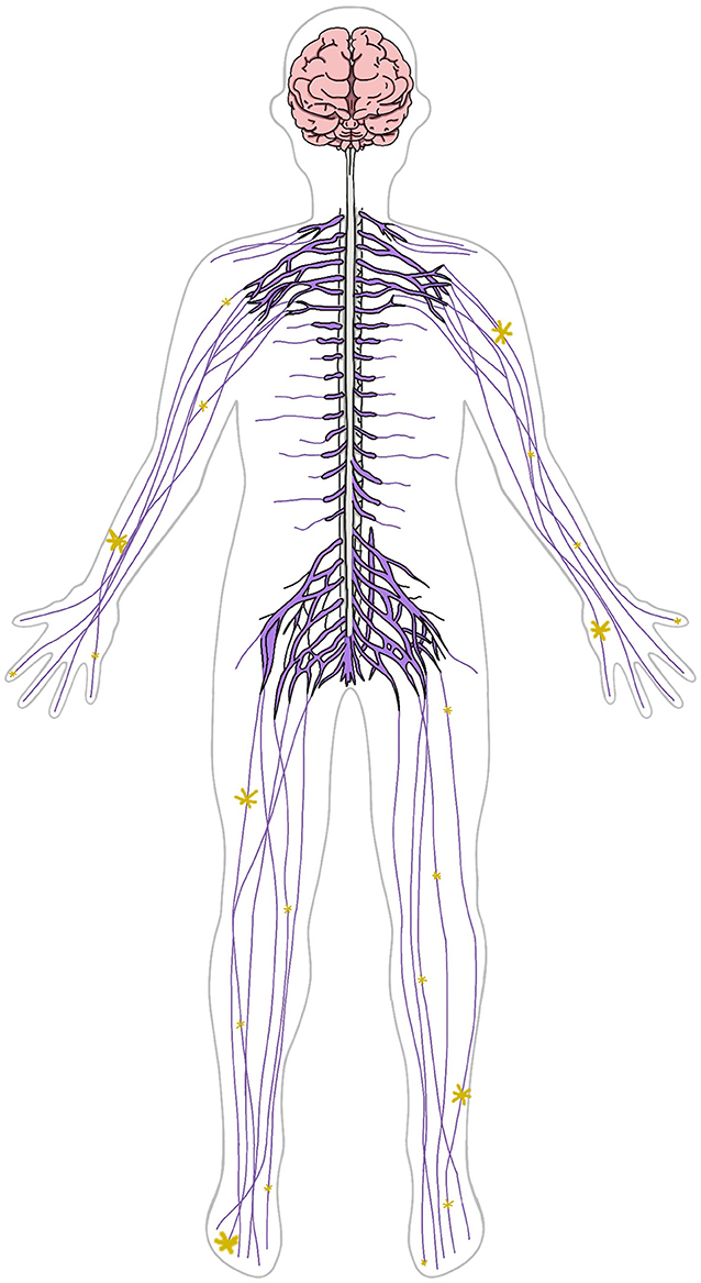
- Figure 1 - The brain is connected to the spinal cord and nerves (purple).
- The yellow stars represent electrical activity, which is carried by the nerves. This electrical activity enables communication between the brain and the other body parts.
When you touch something, like a soft blanket or a hot stone, a message is sent to your brain through the nerves. Your brain then gets the information and tells you, “Hey, that is a soft blanket!” or “Ouch, that is hot! Take your hand away!”.
Neurons also help us move our bodies. If you decide to move your hand, for example, your brain sends a message via your nerves to the muscles in your hand. A different set of nerves will let you know that your hand is actually moving. That means that messages can go in both directions: away from the brain and toward the brain.
Neurons Talk To Each Other
The “arms” or extensions of two nerve cells are connected via switch points called synapses. As you just learned, information travels along the neurons in the form of electrical activity. Once the information reaches the end of one neuron, it must cross the synapse to get to the next neuron. The electrical activity changes to chemical activity to allow the information to pass from one neuron to another.
The brain consists of more than 80 billion neurons, and they all manage to communicate with each other in an organized and effective way. When neurons communicate with each other, it results in electrical brain waves that can be measured by scientists or doctors. Sometimes brain cells chat with each other in a calm and slow way, but other times they are excited and brain activity is fast. Brain waves reflecting that fast brain activity are called high frequency oscillations.
How Can We Measure Brain Activity?
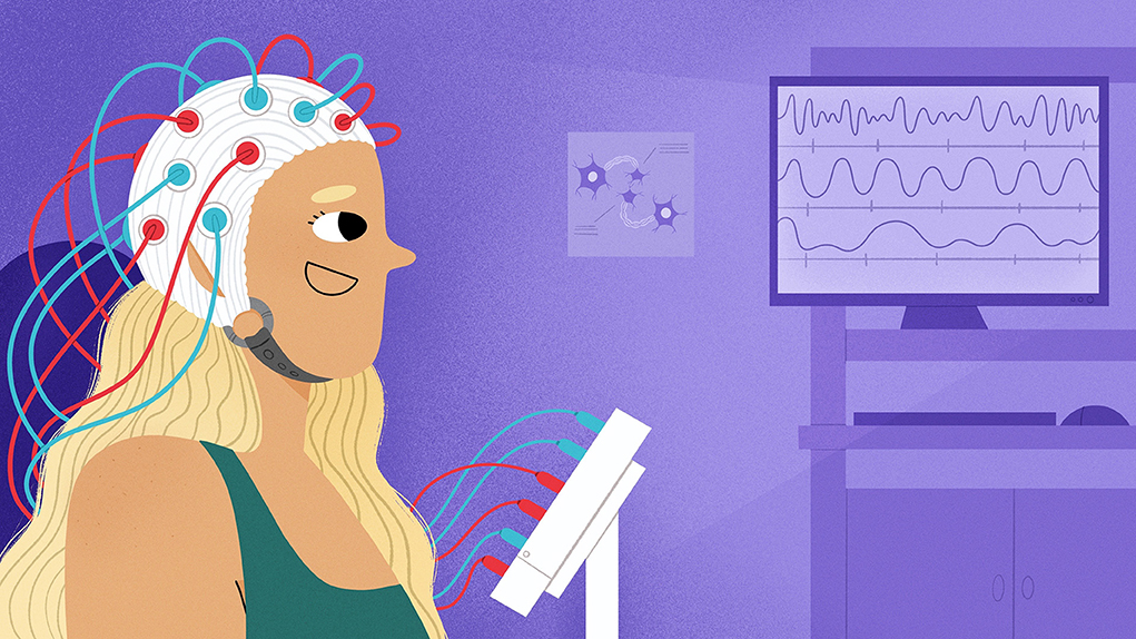
Electrical activity can be measured by a test called electroencephalography (EEG). For EEG, scientists or doctors place tiny metal sensors called electrodes on a person’s head (Figure 2A). The electrodes can detect the small electrical signals produced by the brain, so it is a bit like having lots of mini-microphones on your head! Those metal electrodes (they all together create a metal cap, which can be seen in Figure 2A) can measure change in electric current and send the signal along wires to a computer. If you want to read more about EEG, there is another great article in Frontiers for Young Minds about this method.
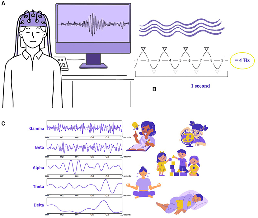
- Figure 2 - (A) A person is wearing an EEG cap with electrodes.
- Wires connect the cap to a computer, where brain waves can be seen on the screen. On the screen, you can see an HFO. (B) Brain waves look a bit like ocean waves. If you draw a horizontal line through the middle of the wave, you can count how many times the wave crosses the line in 1 second. The frequency of this wave is 4 Hz. (C) Brain waves are characterized by their frequency and named after Greek letters. Each kind of brain waves occurs during specific activities, shown on the right.
The EEG activity is displayed on the computer screen and looks like waves going up and down, like the waves in the ocean. The faster the brain works, the faster the waves are. Researchers can even tell if the electrodes are measuring a higher signal on the left or right side of the brain.
The speed of waves is called their frequency, which is measured in a unit called Hertz (Hz). If you draw a horizontal line through the middle of the wave, the frequency is essentially how often the wave crosses that line (Figure 2B). You can see that the wave line goes 9 times past the middle line. For the frequency, you only count the top peaks of the wave (black arrows) within 1 second.
Brain waves are named according to their frequency using letters from the Greek alphabet (Figure 2C). Slower waves are called delta and theta waves, and they mostly occur when the brain is sleeping. Theta waves can also occur when people daydreaming. In healthy children, theta waves can also occur while they are awake. Faster alpha waves can be seen when people are awake and relaxed. Beta waves are dominant in those who are alert or anxious or who have their eyes open, but we can also see beta waves during sleep, when vivid dreams are happening. In this article, we will focus on super fast waves, called high frequency oscillations (HFOs). HFOs are 10 times faster than the usual brain activity (between 80–150 Hz) and can be a sign of the brain working really hard or being in trouble [1, 2].
When Do We Need Super Fast Brain Activity?
HFOs have been seen by EEG whenever people are engaging in a very focussed activity that requires many brain areas to work together with high precision. Two types of computer games are great examples of when our brains use HFOs. First, games that requires fast finger movements and quick reactions, like fighting, jumping, or race car driving, cause the brain to use HFOs to plan movements. Second, HFOs are also needed to memorize locations and activities in more complicated games. Have you ever lost track of where you are in an imaginary world, or forgotten where the villain is hiding? If not, you can thank your brain for doing a great job storing your memories (Figure 3). Storing memories largely happens during sleep. HFOs help to transport memories between parts of the brain that allow you to know your way around in a computer game—not only the same day, but even weeks later [3–5].

- Figure 3 - Illustrates an important brain function: On the right, you can see the same screen after placing trees, plants and houses.
- To be able to look on the left screen and remember in which part the house, trees, plants and birds were placed, your brain had to do the job storing memories before.
When The Brain Gets Too Active
You just learned that fast brain activity can be super helpful in our daily lives. However, sometimes neurons start communicating too fast or in a way that is less coordinated than usual. This can happen in adults and kids who have epilepsy. Epilepsy is a condition in which people experience a sudden loss of control over their body functions because their body parts are not receiving the proper information. These events are called epileptic seizures. During a seizure, the body starts to move uncontrollably or the person experiences a feeling, a scent, or a lack of awareness (almost like daydreaming) for a brief moment. Epilepsy is a fairly common disease, affecting around 1% of children. This means that if you have 500 kids at your school, it is likely that 4–5 of them have epilepsy.
EEG is a very useful test when scientists or doctors want to learn about brain activity in individuals who have seizures. EEG can tell us when the brain’s electrical activity is out of order. Recent research has shown that HFOs can be seen in the EEGs of patients with epilepsy. In contrast to the HFOs that help with thinking, planning, and memorizing, we believe that epileptic HFOs are a sign of the brain being overactive and out of control. This is exciting news because measuring HFOs using EEG can help us to better understand a patient’s epilepsy [6]. HFOs have helped doctors to predict which kids might only have one seizure and which might have epilepsy and be prone to more seizures. It can also help doctors to know whether a person needs medication for their epilepsy and whether that medication is successful in treating seizures [6].
HFOs can also help scientists and doctors to learn which parts of the brain are causing epileptic seizures. This is really important, as some epilepsy patients only have seizures from small regions of their brains. Surgery to remove the seizure-causing brain region can cure these patients (see this Frontiers for Young Minds article for more information) [7]. We think that HFOs are what is called a biomarker for epilepsy. This means that measuring HFOs can help doctors to know how active the disease is and how to successfully treat it.
What Did You Learn?
The brain is like a super storage unit for all the things you have ever experienced. It remembers your favorite moments, like birthdays, family vacations, or adventures with your friends. You have learned that the brain also controls the body’s movements, directing all the different body parts to work together in harmony. HFOs help the brain to function properly and engage into focussed activity that requires many brain areas to work together. However, if HFOs get out of control, a seizure can result. HFOs are an exciting biomarker that can help researchers and doctors to understand how the brain works and help them to diagnose and plan treatments for patients with epilepsy.
Glossary
Neurons: ↑ Nerve cells with bodies located in the brain and long extensions going into different parts of the body. Their job is to pass along important messages helping your body do things like move, think, and feel.
Synapses: ↑ Small gaps between neurons that information must cross to travel from one neuron to another.
Electroencephalography (EEG): ↑ A method to look at the brain’s electrical signals using tiny sensors called electrodes, which send the information to a computer where it can be analyzed.
Frequency: ↑ A measure to determine the speed of waves. It is measured in a unit called Hertz (Hz).
High Frequency Oscillations (HFOs): ↑ Superfast brain waves caused by excessive and sometimes uncoordinated electrical activity inside the brain.
Epilepsy: ↑ A condition of the brain in which a person is prone to having epileptic seizures.
Epileptic Seizure: ↑ A sudden burst of electric activity in the brain, which can cause loss of control of the body or parts of the body.
Biomarker: ↑ A clue that helps doctors understand how the body is doing. HFOs as a biomarker can help to know how active the disease is and how to successfully treat it.
Conflict of Interest
The authors declare that the research was conducted in the absence of any commercial or financial relationships that could be construed as a potential conflict of interest.
Original Source Article
↑Jacobs, J., Staba, R., Asano, E., Otsubo, H., Wu, J. Y., Zijlmans, M., et al. 2012. High-frequency oscillations (HFOs) in clinical epilepsy. Prog. Neurobiol. 98:302–15. doi: 10.1016/j.pneurobio.2012.03.001
References
[1] ↑ Jacobs, J., Schönberger, J. 2019. In search of epileptic scalp high-frequency oscillations. Clin. Neurophysiol. 130:1172–1174. doi: 10.1016/j.clinph.2019.04.006
[2] ↑ Jacobs, J., Staba, R., Asano, E., Otsubo, H., Wu, J. Y., Zijlmans, M., et al. 2012. High-frequency oscillations (HFOs) in clinical epilepsy. Prog. Neurobiol. 98:302–315. doi: 10.1016/j.pneurobio.2012.03.001
[3] ↑ Jacobs, J., Banks, S., Zelmann, R., Zijlmans, M., Jones-Gotman, M., and Gotman, J. 2016. Spontaneous ripples in the hippocampus correlate with epileptogenicity and not memory function in patients with refractory epilepsy. Epilepsy Behav. 62:258–266. doi: 10.1016/j.yebeh.2016.05.025
[4] ↑ Lachner-Piza, D., Kunz, L., Brandt, A., Dümpelmann, M., Thomschewski, A., and Schulze-Bonhage, A. 2021. Effects of spatial memory processing on hippocampal ripples. Front. Neurol. 12:620670. doi: 10.3389/fneur.2021.620670
[5] ↑ Bruder, J. C., Wagner, K., Lachner-Piza, D., Klotz, K. A., Schulze-Bonhage, A., and Jacobs, J. et al. 2022. Mesial-temporal epileptic ripples correlate with verbal memory impairment. Front. Neurol. 13:876024. doi: 10.3389/fneur.2022.876024
[6] ↑ Jacobs, J., LeVan, P., Chander, R., Hall, J., Dubeau, F., and Gotman, J. 2008. Interictal high-frequency oscillations (80–500 Hz) are an indicator of seizure onset areas independent of spikes in the human epileptic brain. Epilepsia. 49:1893–1907. doi: 10.1111/j.1528-1167.2008.01656.x
[7] ↑ Zijlmans, M., Jacobs, J., Zelmann, R., Dubeau, F., and Gotman, J. 2009. High frequency oscillations and seizure frequency in patients with focal epilepsy. Epilepsy Res. 85:287–292. doi: 10.1016/j.eplepsyres.2009.03.026