Abstract
The os cordis (heart bone) is a rare bone found only in a few animals in the world. We discovered an os cordis in some chimpanzees. The os cordis was found in males and females, and in young and old animals. It was not present in chimps with healthy hearts, only in those with severe heart disease. We also discovered that a tissue called cartilage was present around the bone. The presence of cartilage gives us clues about how and why these rare bones develop.
Why are Chimpanzees Interesting?
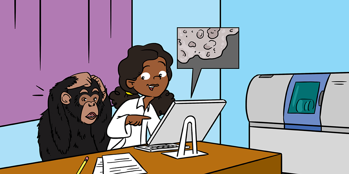
Chimpanzees are our closest living relatives (Figure 1A). We share 95–98% of our DNA with them. Like humans, chimps can use tools, learn sign language, laugh, get angry, kiss, and tickle. Orphaned chimps are often adopted by other chimps. Because of these similarities, we can strongly relate to these animals. Sadly, the big difference between people and chimps is that chimps are endangered. The only wild chimpanzees left today live in Africa. Their rainforest homes are being cut down and they are also hunted for meat. In the wild, chimpanzees live for an average of 15 years, but the oldest lived to be 63. In zoos and sanctuaries, males usually live for nearly 32 years and females live for about 39 years. Like humans, chimps can also get certain diseases. One of the diseases both humans and chimps suffer from is heart disease. We wanted to understand why chimps die of heart disease and what is happening inside their hearts1.
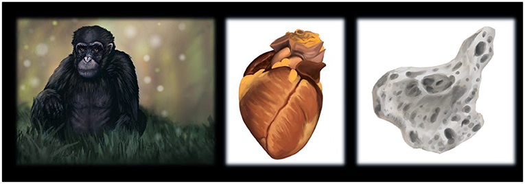
- Figure 1 - Drawings of (A) a chimpanzee, (B) a chimpanzee heart, and (C) an os cordis discovered within a chimpanzee.
- This os cordis measured 7.4 mm in length, 7.6 mm in width and had a depth of 4.2 mm, so it was very small.
The Amazing Discovery of A New Bone
To look inside chimpanzee hearts, we used a high-tech X-ray imaging method called computed micro-tomography, or micro-CT (Figure 1B). This scanner showed the hearts at a very high magnification, which had never before been seen by scientists. We were amazed to discover bones within some of the hearts [1]. The scientific name for this heart bone is os cordis (Figure 1C). We were the first people to see a chimp os cordis, and we were excited about this new finding. To understand as much as possible about this bone, we did further micro-CT scans, looked at the bones and the hearts under the microscope, and analysed the medical histories of the animals.
Which Scientific Techniques Did We Use?
To examine the hearts, each heart was placed into the micro-CT scanner. The heart slowly rotated in the machine and a special camera collected hundreds to thousands of X-ray images from every direction. X-rays are useful because they can detect objects with differing densities [2]. In the hearts, the dense bone looked white on the X-ray image, and the tissue and air appeared grey or black. Each individual X-ray image was sent to a computer and used to compile 3D images of the heart and os cordis. We took exact measurements of each heart and os cordis, and even measured the thicknesses of the delicate bone structures, which the computer represented with different colours in the images (Figure 2A).
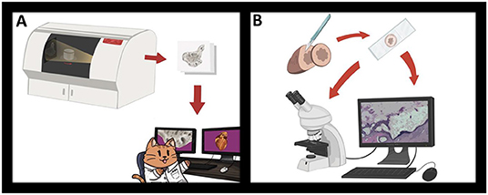
- Figure 2 - We investigated chimpanzee hearts and the os cordis using two main techniques.
- (A) Computed micro-tomography allowed us to discover the bone and understand its structure. (B) Histology allowed us to look at heart and bone cells using staining techniques and microscopes. The images on the cartoon computer screens are real images of the os cordis.
Another important technique that we used is called histology. We removed pieces of the hearts using a scalpel, dehydrated the heart tissue, and surrounded the tissue with paraffin wax. This enabled us to cut very thin slices of the tissue, ten times thinner than a human hair, which we put onto microscope slides. We stained the tissue on the slides with chemicals that make the cells visible under the microscope. Using the microscope and camera, we could see the heart cells, whether the heart had an os cordis, and whether the animals had heart disease (Figure 2B).
We also looked at the ages of the chimps, examined the structures of their hearts, and looked at the medical histories of each animal, to see if they had health problems. This medical information, together with the micro-CT and histology data, helped us to link os cordis to disease.
What Else Did We Discover About the Chimp Os Cordis?
Three out of sixteen of the chimps we studied had an os cordis (Figure 3), and one chimpanzee had cartilage in its heart. Cartilage is a support tissue in the body that is softer and more flexible than bone. We also discovered cartilage tissue around the os cordis bones. This was a fascinating finding because not much is known about how the os cordis grows. Every bone in the body either grows spontaneously or develops from cartilage. The fact that we found cartilage surrounding the os cordis suggests that this bone might develop from cartilage. We also saw blood vessels inside the os cordis. Blood vessels are essential for providing oxygen and nutrients to tissues, including growing bones “Blood vessels under the microscope [3].”
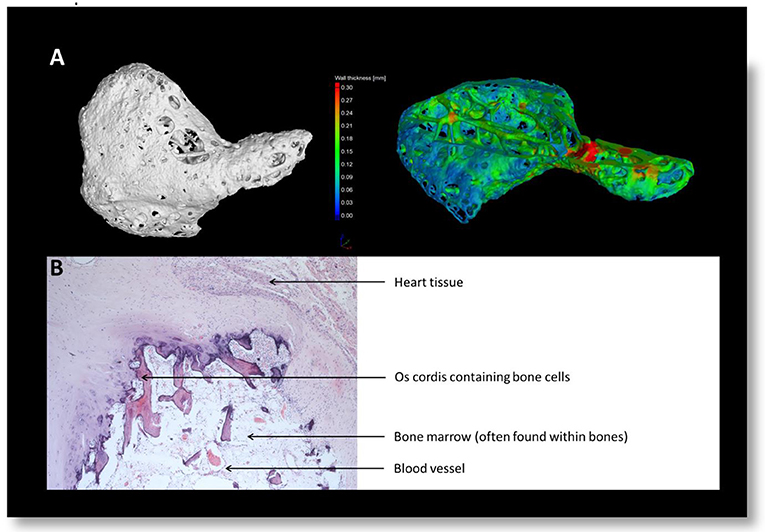
- Figure 3 - Images of the os cordis.
- (A) Using computed micro-tomography, the computer could apply colours showing the thickness of the bone. Thickness ranged from 0.03 to 0.30 mm. (B) Using histology, we could see the cells and blood vessels within the bone using a microscope.
The os cordis was present in both male and female chimps, and in both young and old chimps. That was interesting because, in some species, the os cordis only exists in older animals. We also noticed that the os cordis was more likely to exist in chimps suffering from a type of heart disease. Chimps get many of the same heart disorders we see in humans. One of those is called idiopathic myocardial fibrosis [4]. “Idiopathic” means we do not know the cause or why it happens. “Myocardial” defines something relating to the heart muscle. “Fibrosis” means the thickening and scarring of the heart tissue. Idiopathic myocardial fibrosis can cause death in both humans and chimps.
Why is The Os Cordis In Some Animals But Not Others?
Only a few other animals are known to have heart bones. The os cordis is common in cows, bulls, and water buffalo [5]. This bone is also present in some sheep, goats, camels, and even otters [6], but it has not been found in most other animals. In some animals, the os cordis is present in healthy young animals, but sometimes it only develops in older animals, such as in water buffalo.
There are lots of theories as to why the heart bone might exist. The os cordis may help conduct electricity through the heart. Electrical conduction is what allows the heart to beat. Other scientists have suggested that the os cordis might prevent electrical conduction, though. We do not know yet if either of these ideas is correct. The os cordis seen in the chimpanzees with heart disease could be causing the heart problems but might also be trying to help the heart work.
Another theory is that the os cordis might serve as a sort of support for the heart, helping the heart keep its structure. However, this does not explain why most animals do not have a heart bone. Also, the os cordis can measure less than a centimetre in size, which probably could not help to keep the structure of a large heart.
Some scientists have suggested that the heart bones may have been more common thousands of years ago, but over the course of evolution, they have become less necessary. Another theory is that the continuous stress the heart is under may cause the heart bones to develop, which may help explain why heart bones are sometimes more common in older animals. There are many more theories as to why the os cordis develops, and there are probably additional theories that we have not even thought about yet!
Conclusion
In summary, our research revealed that some chimps have a heart bone, especially if they have heart fibrosis. Both male and female and both young and old chimps can have an os cordis. We also discovered that cartilage was present around the bones, indicating that the os cordis may develop from cartilage. Our research has helped us to better understand the os cordis, chimp hearts, and heart disease.
Just days after we published our scientific paper, another very exciting paper reported that some people have bone cells in their hearts [7]. As with a lot of research, our discoveries leave some big questions unanswered. How and why does the os cordis develop? Why are the heart bones in some species and not others? Why are they sometimes present in older animals or those with heart problems, but not in young, healthy hearts? We still have more research to do in the future to help uncover the answers. Finding a new bone in a species is a rare event and an exciting scientific discovery. It brings up the question of whether there are other animals, including some humans, that could have additional mysterious bones that are yet to be discovered!
Glossary
Computed Micro-tomography (Micro-CT): ↑ An imaging technique that uses x-rays to visualise objects in 3D, at very high magnification.
Os Cordis: ↑ A bone that develops in the heart.
Histology: ↑ A field of study in which microscopes are used to investigate the small structures and cells of the body.
Cartilage: ↑ An important tissue in the body that provides support but is softer and more flexible than bone.
Idiopathic Myocardial Fibrosis: ↑ A heart disorder without a known cause that causes thickening and scarring of heart tissue and can result in early death.
Conflict of Interest
The authors declare that the research was conducted in the absence of any commercial or financial relationships that could be construed as a potential conflict of interest.
Acknowledgments
The authors would like to thank the Anatomical Society for helping to fund this work a Public Engagement and Outreach grant to Catrin titled Anatomy for ALL—Making Anatomy Accessible. We would also like to thank the British Science Association and University of Nottingham for awarding Catrin with a BSA Media Fellowship 2019. We would like to thank all of the authors of the original paper Moittié, S., Baiker, K.,Strong, V., Cousins, E., White, K., Liptovszky, M., Redrobe, S., Alibhai, A., Sturrock, C.J., and Rutland, C.S. This was a collaboration between University of Nottingham, Twycross Zoo and the Ape Heart Project.
Footnote
1. ↑If you are interested in learning more about hearts, see our previous Frontiers for Young Minds article https://kids.frontiersin.org/articles/10.3389/frym.2018.00019
Original Research Article
↑Moittié, S., Baiker, K., Strong, V., Cousins, E., White, K., Liptovszky, M., et al. 2020. Discovery of os cordis in the cardiac skeleton of chimpanzees (Pan troglodytes). Sci Rep. 10:9417. doi: 10.1038/s41598-020-66345-7
References
[1] ↑ Moittié, S., Baiker, K., Strong, V., Cousins, E., White, K., Liptovszky, M., et al. 2020. Discovery of os cordis in the cardiac skeleton of chimpanzees (Pan troglodytes). Sci Rep. 10:9417. doi: 10.1038/s41598-020-66345-7
[2] ↑ Keane, M., Paul, E., Sturrock, C. J., Rauch, C., and Rutland, C. S. 2017. “Computed tomography in veterinary medicine: currently published and tomorrow’s vision,” in Computed Tomography–Advanced Applications, ed A. M. Halefoglu (London: InTechOpen). p. 271–89.
[3] ↑ Machado, M. J., Mitchell, C. A., Franklin, J., Thorpe, A., and Rutland, C. S. 2020. Blood vessels under the microscope. Front. Young Minds. 8:151. doi: 10.3389/frym.2019.00151
[4] ↑ Strong, V. J., Martin, M., Redrobe, S., White, K., and Baiker, K. 2018. Retrospective review of great ape cardiovascular disease epidemiology and pathology. Int. Zoo. Yearb. 52:113–25.
[5] ↑ James, T. N. 1965. Anatomy of the sinus node, av node and os cordis of the beef heart. Anat. Rec. 153:361–71.
[6] ↑ Egerbacher, M., Weber, H., and Hauer, S. 2000. Bones in the heart skeleton of the otter (Lutra lutra). J. Anat. 196 (Pt. 3):485–91. doi: 10.1046/j.1469-7580.2000.19630485.x
[7] ↑ Trainini, J., Lowenstein, J., Beraudo, M., Wernicke, M., Trainini, A., Llabata, V. M., et al. Myocardial torsion and cardiac fulcrum. Morphologie. (2020) 105:15–23. doi: 10.1016/j.morpho.2020.06.010