Abstract
Have you ever wondered how doctors look inside a person’s heart without opening their chest? The heart is a hard-working organ, and thanks to modern technology, doctors can explore it in incredible detail. Several common methods, ranging from classic X-rays to techniques that use strong magnets or sound waves to create images of the heart, can show doctors how a person’s heart beats, pumps blood, and keeps them alive. The coolest part? Some methods let doctors not only see the heart but also fix problems on the spot. Each imaging technique has its own special strengths, allowing doctors to choose the best option for each unique case. With these amazing tools at their fingertips, doctors can understand and care for patients’ hearts like never before.
The Structure of the Heart
When asked to visualize a heart, what do you see? Chances are, you might think of the bright red heart emoji common on social media. But what about doctors, who have studied the organ extensively for many years and seen it in all of its real-life shapes and forms? From their point of view, the heart is a biological machine that never takes a break, pumping oxygen- and nutrient-carrying blood throughout the whole body. This special organ is comprised of 4 chambers: 2 smaller ones called atria on the top, and 2 larger ones called ventricles on the bottom (Figure 1). These chambers work together, contracting in a well-coordinated rhythm to keep blood moving smoothly. Blood circulates into the heart through the atria, then down to the ventricles. It first passes through the right side of the heart, to reach the lungs and refill on oxygen. Then, it returns to the left side of the heart and leaves through a large blood vessel, called the aorta, to nourish the entire body, including the heart itself.
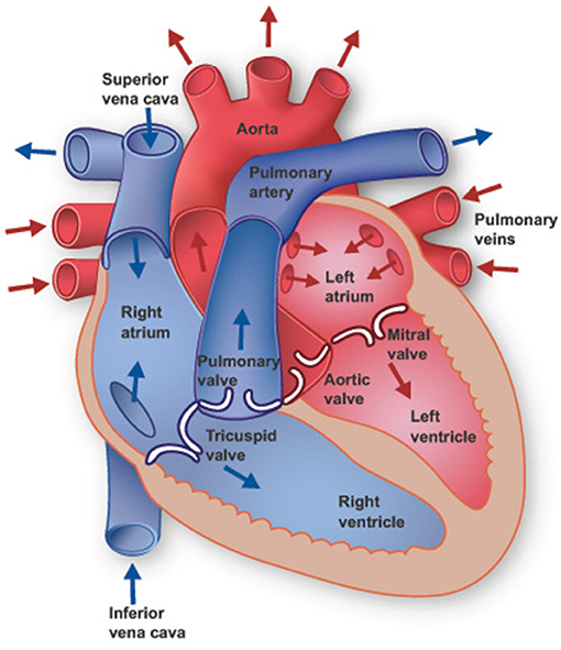
- Figure 1 - The heart has four major compartments, two atria and two ventricles.
- Arrows show the normal blood flow going in and out of the heart (figure credit: The Texas Heart Institute).
What Can Go Wrong With the Heart?
When a patient sees a doctor because of discomfort, pain, or a heavy feeling in their chest, there are many things for the doctor to consider. The discomfort could be caused by something called an arrhythmia, which is a problem with the way the heart is beating. Arrythmia can lead to feelings of a racing heart, called palpitations, and can change the proper flow of blood. If the coronary arteries (the vessels supplying blood to the heart) are blocked by build-ups of fat and other substances—a condition called atherosclerosis—this could cause pain and other heart problems [1]. The heart’s valves are also crucial because they act like gates to ensure blood flows in the right direction. When the valves are defective because of an infection or some other reason, the blood can flow back into the heart instead of moving through the body.
The symptoms a patient experiences might not even stem from the heart at all—they may instead come from the lungs or stomach. To properly diagnose what is wrong with patients, doctors use many special imaging tools and technologies to look inside the body and see what is going wrong.
The Age-Old X-ray and Its Younger Brother CT
Two staples of imaging technology are X-rays and computed tomography (CT) scans, and they are both great choices for examining the heart and lungs. X-rays work by sending invisible beams of light through the body. As these beams pass through, some are absorbed by the bones and tissues, while others travel all the way through. This creates a picture on a special plate on the other side of the body, showing what is happening inside! (Figure 2A). Doctors usually take two different X-ray views, one from the back and one from the side, to get a good look at the anatomy of the chest, capturing not only the heart but also the lungs, ribs, and spine. However, X-rays do not always provide enough detail in more complex cases.
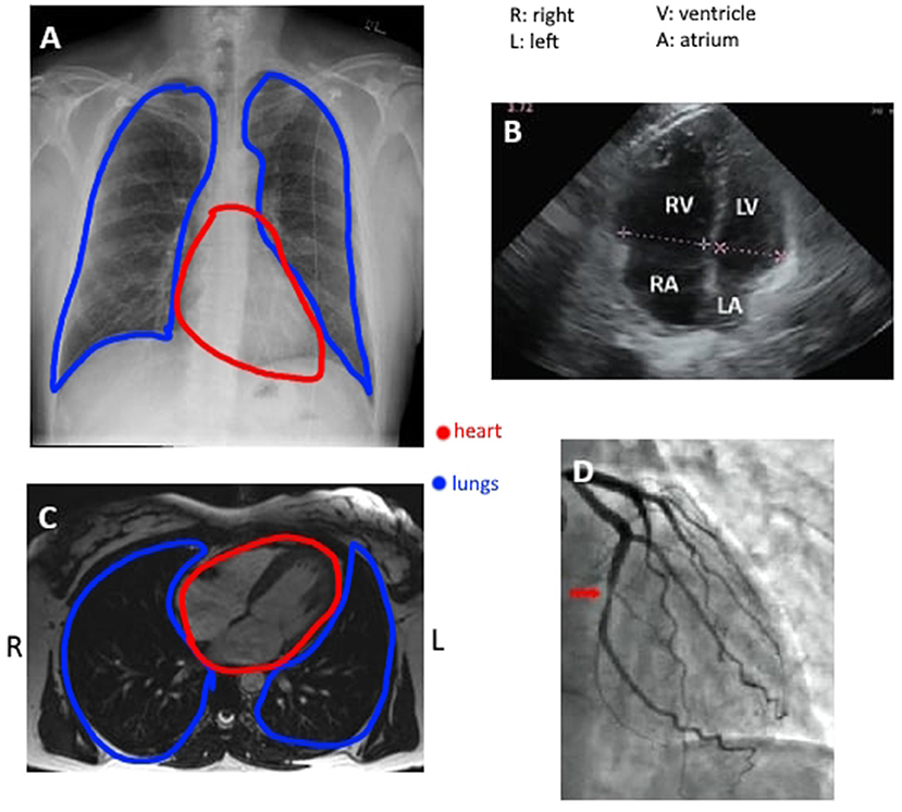
- Figure 2 - (A) A standard view of the chest using an X-ray, where the heart can be seen in the center, leaning slightly to the left.
- (B) A heart image captured by ultrasound, with the major compartments identified by their initials. (C) View of the heart using MRI, from bottom up. (D) Angiography shows the blood vessels that surround and nourish the heart. Notice how each tool focuses on a different aspect of the heart (figures from Wikimedia Commons).
That is where CT scans come in handy—they are like a new and improved version of X-rays. CT scans take multiple X-ray images at different angles all around the body and combine them to create a detailed 3D model of the heart. The resulting 3D image is divided into multiple sections, like slices of a bread loaf, that clearly show the blood vessels throughout the heart, making CT an excellent test to see blockages caused by atherosclerosis [2]. Doctors can also inject a special dye, called contrast, to make the heart’s structures stand out even more clearly. While CT has impressive imaging capabilities, it also has downsides. For one, because of the number of X-rays needed to build the 3D image, CT scans expose patients to a considerable amount of radiation. The dosage is usually harmless for the average healthy adult. However, for a child, CT scans can be much more problematic because they can increase the risk of cancer many years after the exposure. To achieve the best image, the scanner also requires the patient to stay relatively still and hold their breath for some time. Nevertheless, the technology is an excellent and accurate tool that doctors around the globe use every day.
Learning from Bats: Ultrasound Imaging
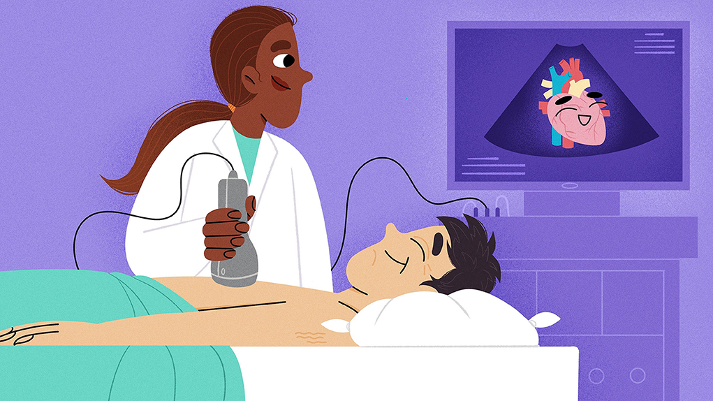
One of the most convenient methods of heart imaging is echocardiography, which provides a live view of the heart, allowing doctors to watch the heart beating in real time. This long word can be divided into 3 smaller words: “echo”, which means sound; “cardio”, which comes from the Greek word for heart; and “graphy”, which means writing or drawing. Similar to how bats navigate in the dark by listening to the echoes of the high-pitched sounds they chirp out, a small handheld device called a transducer sends out ultrasound waves that bounce off the tissues in the body. Harder surfaces, like bone, reflect more waves, while softer tissues, like blood, reflect fewer. The machine processes this feedback to create the image the doctor can see on the screen, which refreshes constantly—up to a 100 times per second [1]. On the ultrasound image, hard surfaces appear white because they reflect more waves, while liquids like blood appear black due to the lack of reflected waves (Figure 2B). Ultrasound is quite useful for visualizing heart structures, such as the valves and chambers, and it can also measure how blood is flowing through the heart by detecting its speed. Usually the transducer is placed on the chest, but if the view is not clear enough, the doctor can image the heart by inserting the device down the patient’s throat, into the esophagus. While this can give doctors a better view, it requires using anesthesia—medication that numbs the patient’s throat. Another important advantage of echocardiography is that it is not harmful to the patient since it does not use radiation like X-rays and CT scans. Echocardiography is a popular choice when doctors need to assess patients urgently, like after traumatic accidents, especially to check whether fluid or blood is building up around the heart.
The New(er) Kid on the Block: MRI
Next is another key imaging method in the world of medicine: magnetic resonance imaging (MRI). As the name suggests, this technology uses a magnetic field to create very detailed images. MRI can identify most structural and functional problems in the heart, like inflammation, blood flow disruptions, or when the heart itself is not getting enough blood (Figure 2C) [3]. An MRI scan also allows doctors to see the aorta, which helps them check for dangerous conditions like aneurysms, which can be very serious. This way, they can make sure everything is healthy and address any problems quickly. One of the main benefits of MRI is that it uses a magnetic field instead of radiation, making it safer for patients. However, this also means that no metal can be within the patient, including pacemakers and metal replacement body parts. Another big challenge is its high cost. To get the best images, a scanner with a very strong magnetic field is needed and not all hospitals and imaging centers can afford these. Its high cost makes MRI less accessible and available than other imaging techniques. Nevertheless, these inconveniences are becoming less of a problem as technology advances [3].
The Old Reliable: Angiography
Last but not least, the gold standard in heart imaging is angiography. During angiography, doctors insert a small tube called a catheter all the way from a blood vessel in the wrist or leg and guide it to the heart [1]. Using an uninterrupted series of X-rays, doctors can get a live view of the coronary vessels around the heart and see how blood is flowing (Figure 2D). They do this by injecting a special dye that the X-ray machine can easily detect, which helps them spot any problems. Angiography can also measure blood pressure levels in each compartment of the heart. Because angiography provides doctors with direct access to the heart and its vessels through the catheter, this method can also be used to repair problematic heart valves. Its double purpose makes it a valuable tool in urgent care situations [4]. Sometimes, angiography can be combined with other methods like CT or echocardiography, to help guide the procedure. The biggest disadvantage of angiography is its increased risk of injuries, such as bruising or damage to the patient’s blood vessels. Also, any dye used in medical procedures can cause problems in people who have weak kidneys, because the kidneys need to work extra hard to flush the die out of the body in the urine. Despite these disadvantages, angiography remains an excellent choice of imaging technology because it creates very good images of the coronary arteries and can be used for both imaging and treatments.
The Whole Package
All in all, it is fascinating that the medical field has developed so many different ways to look at a single organ. Thanks to the help of all the techniques we discussed, the health field has learned so much more about the heart and its mechanisms. As technology progresses, current methods will get better at overcoming their limitations, becoming more affordable and efficient, and completely new approaches will probably be developed, too. Even now, there are many other cardiac tests that are outside the scope of this article, from variations of the techniques we discussed to entirely different technologies. Examples include performing imaging tests during exercise to assess the heart’s response to stress, electrocardiograms that measure the heart’s electric signals, or even nuclear scans using radioactive markers [1]. Healthcare professionals have an arsenal of tools at their disposal to examine the heart, but while it is great to have many options, it can also be difficult to find the best choice for each scenario. Heart specialists need to use their knowledge and experience to carefully balance the pros and cons of each tool, to avoid missing or delaying a diagnosis. With all these tools and continuing advancements, doctors are better equipped than ever to understand and care for our hearts.
Glossary
Aorta: ↑ The biggest artery in the body. All blood leaving the heart to supply the body with oxygen and nutrients passes through this blood vessel.
Arrhythmia: ↑ An abnormal heartbeat, which can be slow, fast, and/or inconsistent.
Coronary Arteries: ↑ Blood vessels surrounding and supplying the heart with oxygen and various nutrients.
Atherosclerosis: ↑ Disease where accumulation of fats and other substances block blood vessels to various degrees, disrupting blood flow.
Imaging: ↑ Process that allows doctors to see inside our body through pictures.
Ultrasound: ↑ Sound waves that are at a much higher frequency than those that we hear, which doctors can use to create an image.
Esophagus: ↑ Part of the digestive tube in the chest, allowing passage of food from the throat to the stomach.
Aneurysm: ↑ Dilation or bulging of a blood vessel at a specific weak point, which can burst if it is severe enough.
Conflict of Interest
The authors declare that the research was conducted in the absence of any commercial or financial relationships that could be construed as a potential conflict of interest.
References
[1] ↑ Rehman, R., and Makaryus, A. N. 2020. Cardiac Imaging. Treasure Island (FL): StatPearls Publishing. Available online at: https://www.ncbi.nlm.nih.gov/books/NBK448128/ (accessed October 28, 2024).
[2] ↑ McKavanagh, P., Walls, G., McCune, C., Malloy, J., Harbinson, M. T., Ball, P. A., et al. 2015. The essentials of cardiac computerized tomography. Cardiol. Ther. 4:117–29. doi: 10.1007/s40119-015-0052-0
[3] ↑ Rajiah, P. S., François, C. J., and Leiner, T. 2023. Cardiac MRI: state of the art. Radiology 307:e223008. doi: 10.1148/radiol.223008
[4] ↑ Omeh, D. J., and Shlofmitz, E. 2021. Angiography. Treasure Island (FL): StatPearls Publishing. Available online at: https://www.ncbi.nlm.nih.gov/books/NBK557477/ (accessed October 28, 2024).