Abstract
When you go to buy a pet fish, you will probably get very detailed instructions on how to take care of it. Even before you go home with your new buddy, you will know a lot of useful stuff, like what it eats and how often you need to clean its tank. Now, if you tried to adopt an octopus, things would not be so easy. I mean, does it even have a mouth? Scientists have a similar problem. When we plan experiments using animals, we need to know a lot about them so that we can tell whether or not our experiments are affecting them. Since scientists cannot hope to learn everything about every animal, they decided to study just a handful of them and use those well-studied examples for their research. These well-studied creatures are called model organisms and, in this article, you will learn about the smallest of them.
Introduction
There is such an incredible variety of living beings that trying to learn everything about all of them would take us hundreds and hundreds of years. Scientists who want to use living beings for research do not want to wait that long—I mean, who is going to win all those Nobel prizes in the meantime? So, instead of studying every type of fish in the sea, we study just a few of them. This principle is particularly important for bacteria, because there are more bacterial species than fish in the sea. In fact, there are so many bacterial species that we can only guess how many there are. And then, to make things even more difficult, bacterial species are so different from each other that sometimes comparing two species feels more like comparing a jellyfish with a horse. Because we know so little about the different kinds of bacteria that exist, the bulk of research done using bacteria has been performed using just a handful of microorganisms called model organisms [1]. Scientists use them when they want to do complicated studies because, since we know a lot about them, it reduces the amount of uncertainty that we could encounter in our experiments. We have learned so much about some model organisms that it is now easy to use them to do the type of experiments we want to do [2].
Bacteria as a Model Organism
Bacteria, just like all other living things, are made of a bunch of chemicals. While there are many kinds of chemicals inside them, two of the most important are DNA and proteins. DNA molecules hold the genetic information of an organism. This genetic information is what gives living organisms their identities—stuff like the color of flowers or the shape of a person’s eyes. The DNA regions responsible for these and many other functions are called genes.
Proteins are also chemicals, but their functions are more diverse. While DNA is like a list of recipes, the proteins are the actual cakes. Proteins come in various forms and sizes and can perform many different functions, but the important thing to know is that they are coded in the DNA, in those genes that I have told you about, and if an organism suffers changes in its DNA sequences, the proteins can be changed in significant ways, sometimes for the better, other times for the worse.
Every organism has DNA and proteins, but what makes bacteria special is that they do not have a lot of these molecules. Well, they do have thousands of genes and proteins, but that is a relatively low number compared with the hundreds of thousands of genes and proteins that animals or plants have. Because of their relative simplicity, it is a little easier to understand the functions and interactions of bacterial genes and proteins. The low numbers of proteins and genes also means that bacteria are generally simpler organisms. While other organisms might have organs dedicated to respiration or food digestion, bacteria do everything at once in their one organ: the cell. Each bacterial cell is a whole organism in and of itself.
Another advantage of working with bacteria is that they do not require a lot of food or space. Due to their small size, they are easily fed so we can maintain whole bacterial populations for days in very small amounts of what is basically chicken broth. Finally, bacteria grow rapidly by dividing themselves into two new cells, so they grow exponentially, which gives scientists a lot of cells to work with.
Here are a few bacterial model organisms that are used for scientific investigations around the world:
Escherichia Coli
E. coli is the undisputed bacterial superstar of the model microorganisms. E. coli is used for almost everything, from studies of the bacterial life cycle to experiments on the way bacteria behave in extremely cold temperatures. This small and round bacterium (Figure 1a), discovered by doctor Theodor Escherich in 1885, has been used in many experiments that helped us to understand how bacteria work: how they eat, how they reproduce, questions about their genes and their proteins—about almost everything, really. So, in a way, it can be said that modern microbiology has been built upon E. coli’s “shoulders.” E. coli are usually found in our guts, where they live without harming us, although some E. coli strains are known to cause diarrhea and other gastrointestinal diseases. But do not worry, the strains used in laboratories are not harmful [1].
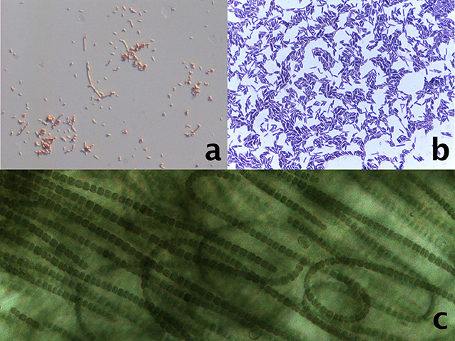
- Figure 1 - Structures of some of the bacteria that are used as model microorganisms.
- The images were taken with a microscopy, because these bacteria are very small. (a) Stained Escherichia coli round cells. (b) Stained Bacillus subtilis cells. (c) The naturally green cyanobacteria Nostoc sp., a bacterium that forms filaments of multiple linked cells.
Bacillus Subtilis
Bacillus is known for the ease with which scientists can manipulate its genes, which allows us to investigate the functions of many of those genes [3]. Another interesting characteristic of this bacterium is that it produces structures called endospores, which are a special cellular form that allows it to survive even when the conditions are not very good for its growth. While Bacillus is not the only organism that can create endospores, most of the studies investigating how endospores form were done in Bacillus [4]. These bacteria are found in the soil and have a rod-like form (Figure 1b), often with endospores found on one end.
Mycobacterium Tuberculosis
These bacteria cause tuberculosis, a disease that used to be very deadly. A lot of the research done using Mycobacterium taught us how to use chemicals that can kill dangerous bacteria. Although tuberculosis is not nearly as deadly as it once was, there are now drug-resistant Mycobacterium strains. These strains are dangerous, because they can survive in the presence of many antibiotics. Currently, much of the research done using Mycobaterium is focused on learning how it infects humans, how the bacteria interact with antibiotics, and how we can defend ourselves from them [5]. Another research field that uses Mycobacterium is the study of bacterial communities. Bacterial communities are bunches of individual Mycobacterium cells attached tightly to each other by a special chemical compound produced by the bacteria, called mycolic acid. These bundles of Mycobacterium cells look like cords, with individual cells wrapping around each other in a disordered way [6].
Streptomyces
You probably know of antibiotics as substances that kill bacteria… but did you know that some bacteria actually produce antibiotics? Streptomyces are great antibiotic producers. For about 20 years, these bacteria were intensively studied and used to produce many new antibiotics. Thanks to that work, we now know much more about how antibiotics are made and about the genes and proteins that are involved [7]. Streptomyces have also been used to study how bacterial cells develop. These bacteria can produce specialized cells called spores, along with long, branching, filaments that sprout out of the cells, which are called hyphae [8]. Those structures give Streptomyces a unique look—the cells are elongated, with branching hyphae that occasionally have round spores around them, making the cells look like colored splashes (Figure 2), due to the array of chemicals that they produce. Streptomyces can be found living in many terrestrial habitats.
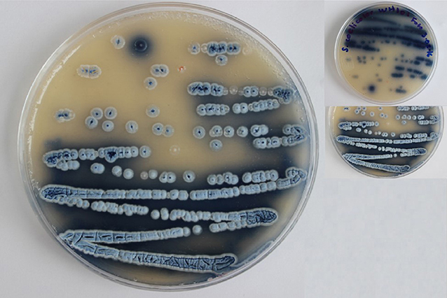
- Figure 2 - A culture of the blue colored Streptomyces coelicolor with white spores.
- They have a particular smell, like a wheat field after it rains.
Cyanobacteria
Cyanobacteria are actually made up of a whole group of related bacteria, which is called a phylum. Within a phylum, all organisms share some characteristics and, in the case of cyanobacteria, the shared characteristic is their lively green color. Their great green look is caused by a protein called chlorophyll, which allows Cyanobacteria to perform photosynthesis. Photosynthesis is a process that has mostly been studied in plants. Like plants, Cyanobacteria can use photosynthesis to change solar energy into chemical energy, which they then use to power themselves [9]. Scientists have studied the proteins and genes in Cyanobacteria that allow it to perform photosynthesis and, in recent years, there has been even more interest in Cyanobacteria as people think about renewable energy. There are now a lot of studies attempting to learn how to harness the photosynthetic potential of Cyanobacteria for industrial applications [10]. Cyanobacteria can be found pretty much everywhere, and many Cyanobacteria species have unique structures (Figure 1c), but all of them are green, thanks to their chlorophyll.
Conclusions
There are a lot of model microorganisms out there, each with unique biological characteristics that can used for different types of scientific studies. We have only shown you the tip of the iceberg—there are many more types of bacteria that are used as model organisms and we could not cover them all here. So, do not get angry if we left your favorite bacteria out!
Acknowledgments
Thanks to Francisco Barona-Gomez, Hilda Ramos-Arboites, Alan Yañez-Olvera, and everybody who helped in the whole process of getting this article out there.
Glossary
Bacteria: ↑ Organisms that only have one cell. The singular is bacterium.
DNA: ↑ A chain of four chemicals arranged in different orders. It is used to codify and store the necessary information to make proteins.
Proteins: ↑ The most abundant chemicals in cells. They have various functions and are made of many different chemicals. The instructions to build them are codified in the DNA.
Cell: ↑ The smallest biological sub-unit. It contains DNA, proteins, and many other chemicals.
Conflict of Interest
The author declares that the research was conducted in the absence of any commercial or financial relationships that could be construed as a potential conflict of interest.
References
[1] ↑ Blount, Z. D. 2015. The natural history of model organisms: the unexhausted potential of E. coli. Elife 4:e05826. doi: 10.7554/eLife.05826
[2] ↑ Fields, S., and Johnston, M. 2005. Whither model organism research? Science 307:1885–86. doi: 10.1126/science.1108872
[3] ↑ Borriss, R., Danchin, A., Harwood, C. R., Medigue, C., Rocha, E. P., Sekowska, A., et al. 2018. Bacillus subtilis, the model Gram-positive bacterium: 20 years of annotation refinement. Microb. Biotechnol. 11:3–17. doi: 10.1111/1751-7915.13043
[4] ↑ Errington, J. 2003. Regulation of endospore formation in Bacillus subtilis. Nat. Rev. Microbiol. 1:117. doi: 10.1038/nrmicro750
[5] ↑ Georghiou, S. B., Magana, M., Garfein, R. S., Catanzaro, D. G., Catanzaro, A., and Rodwell, T. C. 2012. Evaluation of genetic mutations associated with Mycobacterium tuberculosis resistance to amikacin, kanamycin and capreomycin: a systematic review. PLoS ONE 7:e33275. doi: 10.1371/journal.pone.0033275
[6] ↑ Zambrano, M. M., and Kolter, R. 2005. Mycobacterial biofilms: a greasy way to hold it together. Cell 123:762–4. doi: 10.1016/j.cell.2005.11.011
[7] ↑ de Lima Procópio, R. E., da Silva, I. R., Martins, M. K., de Azevedo, J. L., and de Araújo, J. M. 2012. Antibiotics produced by streptomyces. Braz. J. Infect. Dis. 16:466–71. doi: 10.1016/j.cell.2005.11.011
[8] ↑ Chater, K. F. 2006. Streptomyces inside-out: a new perspective on the bacteria that provide us with antibiotics. Philos. Trans. R. Soc. B Biol. Sci. 361:761–8. doi: 10.1098/rstb.2005.1758
[9] ↑ Peschek, G. A. 1999. “Photosynthesis and respiration of cyanobacteria,” in The Phototrophic Prokaryotes, 1st Edn., eds G. A. Peschek, W. Löffelhardt, and G. Schmetterer (Boston, MA: Springer), 201–9. doi: 10.1007/978-1-4615-4827-0_24
[10] ↑ Lindberg, P., Park, S., and Melis, A. 2010. Engineering a platform for photosynthetic isoprene production in cyanobacteria, using synechocystis as the model organism. Metab. Eng. 12:70–9. doi: 10.1016/j.ymben.2009.10.001