Abstract
Heart disease is a major cause of health problems worldwide. There are many different types of heart disease, but one that is fairly common and can have lots of negative side effects is called cardiomyopathy. We know that humans and many mammals, including dogs, get cardiomyopathy. In dilated cardiomyopathy the heart changes shape and often the electrical signals within the heart change. Because of these changes, the heart is not able to pump blood around the body very well, which means that oxygen and nutrients are not delivered to the body at the optimal levels. This can affect day-to-day life, and it can also cause early death. Understanding cardiomyopathy and finding the mutations in genes that cause it are essential, as this information can help us to diagnose, treat, and prevent this heart disorder. We can try to mend the broken hearts that do not work properly. In dogs with cardiomyopathy, we have discovered genetic mutations and found links between these changes in the dogs’ hearts.
The Heart
The heart pumps blood to the lungs where oxygen goes into the blood, it is then pumped back to the heart and then this blood gets pumped around the body. This is essential to deliver oxygen and nutrients to every cell in the body and take away waste including carbon dioxide. The heart has two ventricles and two atriums (called atria) which help to pump the blood. The blood enters these chambers and the ventricles and atria contract, pushing the blood into the blood vessels. The importance of the heart has been known for thousands of years. The famous Greek philosopher Aristotle, born 2,402 years ago, said the heart was the most important organ of the body. Plato, another philosopher, and Hippocrates, a great doctor, worked in the same era as Aristotle and they noticed that the heart was important for pumping blood around the body.
The heart has also become a very important symbol of emotions throughout history, in cultures and religions across the world. It is a symbol of love, a place where the soul might live and where emotions and reason come from. We see this symbolism and importance in religions such as Christianity, Hinduism, Judaism, Islam, and many more. From ancient Romans and Aztecs through to modern-day Chinese medicine, Cupid and his arrows, and Valentine’s Day, the heart is a very special symbol. How many times a day do we see it as an emoji? Throughout time and in every part of the world, the heart is a necessary organ and helps us show emotion.
How Do Hearts Get Broken?
Often when we hear the words, “a broken heart,” we think of a person who has lost someone or something they love. There is actually a heart disorder that is referred to a “broken heart syndrome”—also called takotsubo or stress cardiomyopathy. It is a temporary but serious heart problem that is rare and very often disappears within days/weeks. It is not only caused by very high levels of sadness though. Shock, extreme sudden happiness, and even stress can cause this problem. It is one of several different types of “cardiomyopathy,” heart disorders that are seen in humans and animals. Common types of cardiomyopathy are called: (1) hypertrophy; (2) dilated; and (3) restrictive [1].
Sadly, not all of these cardiomyopathies simply disappear with time and most are caused by something other than emotions. Cardiomyopathy can cause the heart to change shape, grow larger than it should (Figure 1), or even start beating incorrectly by changing the cardiac (heart) electrical signaling. Sometimes cardiomyopathy does not affect how a person lives, and they might never know they have heart problems. In other cases, cardiomyopathy can be very dangerous, causing illness and even death if the heart stops beating. The heart can become damaged. Cardiomyopathies affect around 1 person in 500, but for some animals this number can be much larger. There are some breeds of dogs in which half of them have cardiomyopathy. My research team wants to understand why cardiomyopathy happens, so we investigate heart disease by looking at genetics, which is the code found inside most cells in the body which controls how our bodies work.
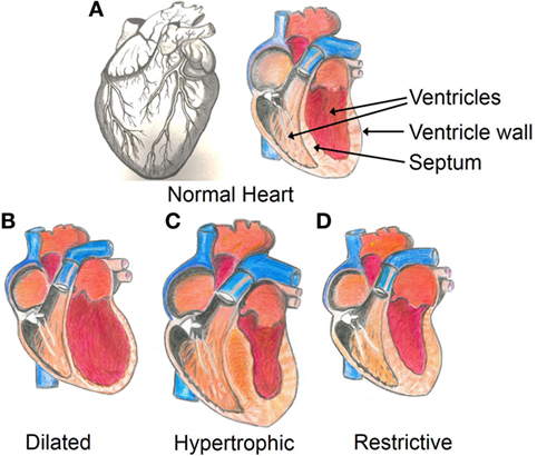
- Figure 1 - Structure of the heart.
- A. Anatomy of a normal healthy heart, from the outside (left) and the inside (right) showing the ventricles which make up the bottom of the heart and the septum separating the left ventricle from the right ventricle. There are different types of cardiomyopathy. B. In dilated cardiomyopathy, the ventricle walls become thinner as the ventricles get larger. C. In hypertrophic cardiomyopathy, the ventricles and/or septum may become thicker and stiffer, and the whole heart can get larger. D. In restrictive cardiomyopathy, the ventricle wall becomes stiff, but not necessarily thicker.
The Hidden Code Behind Heart Disease
To keep a healthy heart, we should all try to be fit, active, eat healthy foods, and avoid lots of alcohol, fatty foods, drugs, and smoking. These actions help to make the heart work as well as it can. Sadly, our genes also play a part in deciding if we are likely to get cardiomyopathy. The genes are the code for life in all plants, animals, humans, and other organisms. Genes exist in nearly every cell in the body and help to run all functions that we need to live and be healthy. Your eye color, hair color (if you do not dye it), height, and even how healthy you are are all partly decided by your genes. Other factors, such as food, chemicals, and your environment play a role too, but the DNA (deoxyribonucleic acid) is the hidden genetic code that helps us be what we are.
Did you know that you have a lot in common with a banana? Over 60% of human DNA is the same as the DNA of a banana; that slug in the garden shares 70% of our DNA, and 95–96% is the same as that of a chimpanzee. We are all a little bit different from each other. Identical twins are natural clones, they start off with the same DNA code as each other, but even they can get mutations throughout their lifetimes. Half of your genetic code comes from your mother and half from your father. Were you aware that your DNA is 99.9% the same as the other humans on our planet? You are nearly the same as Albert Einstein, Rosalind Franklin (who did the experiments that showed the structure of DNA), Stephen Hawking, or even your favorite singer or astronaut.
What is the DNA Code?
The DNA code is fairly simple. Four “bases” called A (adenine), C (cytosine), G (guanine), and T (thymine) join together in pairs—T + A and G + C (Figure 2). The pairs line up to create genes. Humans have around 3 billion base pairs, which we call the human genome. These genes are organized into 46 chromosomes in the human (this number can be different for other species), which are found inside the nuclei of the cells that make up our bodies. Around 20,000 genes create proteins that the body uses to function. If the genetic code changes, which we call a mutation (Figure 3), the proteins made from the code might also change, or the protein might not be made at all. This means that the body might function differently or not function at all. Mutations might be passed on by your parents, but they can also happen spontaneously inside your cells.
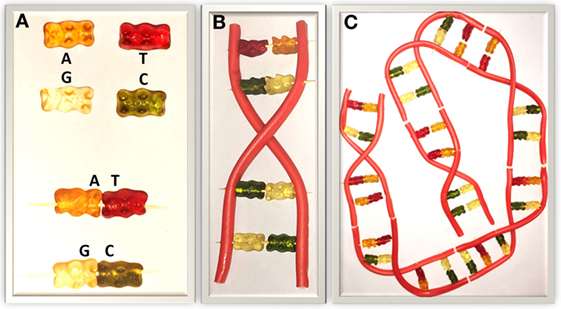
- Figure 2 - Structure of DNA modeled using gummy bears and liquorice.
- A. The four DNA nucleotide bases: A (adenine), C (cytosine), G (guanine), and T (thymine). They are represented by the gummy bears. B. The bases pair up on a structure called the “backbone,” (the liquorice). T + A and G + C bind together. Several base pairs create a gene. C. Many genes together create a chromosome.
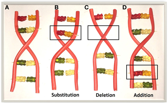
- Figure 3 - DNA mutations using gummy bears and liquorice.
- Examples of some DNA mutations. A. A simple DNA sequence is shown. B. The DNA sequence changes, as the bases swap from a G + C to T + A. This mutation is called a substitution. C. A deletion is when the DNA sequence has lost a base pair. D. An addition is when the DNA sequence has a base pair added. Substitution, deletion, or addition of more than one pair of bases can also happen.
In fictional examples such as the X-Men, DNA mutations cause extraordinary human superpowers—mindreading, blue skin, shape shifting, and flying. In real people, actual mutations might just mean you have blonde hair instead of brown hair, which is obviously not a problem. If this kind of harmless mutation is present in 1% of the population, it is usually called a polymorphism. In other cases, a mutation might mean that a person gets a disorder or syndrome, and there could be negative side effects and health problems. Common examples are Down syndrome, cystic fibrosis, and cancers, which are all caused by mutations in lots of different genes. So far, we have identified over 6,000 genetic disorders, and that number is still increasing. We now know that mutations in lots of genes can cause cardiomyopathy, and we are still searching for more possible mutations.
The Six-Billion-Base Hunt for Disease
Searching through the six billion DNA bases in the genome is not easy. The human genome project was the largest collaborative biological project in the world and included scientists from America, United Kingdom, France, Australia, China, and other countries. It was such an important project that American President Clinton and British Prime Minister Blair jointly announced the publication of the human genome in June 2000. Researchers around the world are still working hard to understand what every gene and mutation does. This work is complicated because each gene or mutation can have many functions, causing different outcomes or even showing up at different times.
Although the basic code of DNA is simple, the specific genes we are born with and how we turn out can be very complicated. Some characteristics or disorders are more likely to show up than others. For example, brown eye color is dominant to blue, so if you have the genes for blue and brown eyes, your eyes will turn out brown. Other characteristics are equally likely to show or may even be expressed together. For example, if you breed a Camellia plant with red flowers with a Camellia plant that produces white flowers, the plants produced have both red and white flowers (not pink flowers, the red and the white are separate). Sometimes, if we have certain codes in two genes at once, we may see a specific outcome, but if we have only one of those codes, we might not see an effect. In one of our research projects, we were able to show that if specific codes were observed in multiple genes, dogs were much more likely to get cardiomyopathy [2]. So, one gene or one mutation alone is not necessarily controlling heart disease. Over the years, scientists have shown that mutations in lots of different genes can cause the same disorder, but we showed that gene combinations are also important in cardiomyopathy [2].
Searching for Mutations in Dogs with Cardiomyopathy
We conducted an international study in which we looked at answering three research questions: (1) which genes are involved in cardiomyopathy; (2) which mutations in those genes could be associated with getting cardiomyopathy; and (3) does having two or more of these mutations affect whether a dog might get cardiomyopathy? Some dogs have very high levels of cardiomyopathy—up to 50% in some breeds [3, 4]. We already know that mutations in over 50 different places on the chromosomes can cause human dilated cardiomyopathy, but there are more mutations to find in people and very few studies have looked at mutations in dogs. We also know that Irish Wolfhounds have a high incidence of dilated cardiomyopathy and have a very short lifespan. The average Irish Wolfhound lives for just 7.04 years, on average. Out of 165 canine breeds, only 14 other breeds live for a shorter period than Irish Wolfhounds, so heart disease is really affecting these dogs.
To study the genes and answer our research questions, we asked dog owners to get some DNA from their dogs by wiping a special cotton swab inside the dog’s mouth. In the laboratory, we were able to extract the DNA from the cells on the swab. We looked at the dogs’ medical backgrounds and diagnosed whether they had cardiomyopathy. A lot of Irish Wolfhounds also get a heart disorder called atrial fibrillation, so we also checked them for this too. The 379 dogs we studied were then classified as (1) healthy with no heart problems; (2) dilated cardiomyopathy only; (3) atrial fibrillation only; or (4) dilated cardiomyopathy AND atrial fibrillation. This was the largest Irish Wolfhound genetic study done in the world. We also looked at males and females separately, because sometimes gender can play an important role in genetic disorders. Then our hunt for mutations began. We looked for single-base mutations in five locations using laboratory techniques called polymerase chain reaction (PCR) and restriction-fragment genotyping. This work involved a team of expert cardiac veterinary surgeons, and scientists who specialize in genetics. After many years, countless clinical checks (seeing if the dogs have heart disease), lots of laboratory studies looking at the DNA, and some biological mathematics, we were able to show the results [5].
We found that 80.5% of the dogs with dilated cardiomyopathy also had atrial fibrillation. There were no differences in the numbers of males and females that had heart problems, but males were more likely to get the symptoms of heart disease at a much younger age. 80% of the males were diagnosed before the age of 6.5 years, but in females the 80% point was reached later, at 8.5 years old. We also discovered that if dogs had one of three specific genetic mutations, they were much more likely to get dilated cardiomyopathy and/or atrial fibrillation. The type of mutation found in those locations also mattered. Different mutations were linked to different risks of getting the heart diseases. Combining different mutations together was even more likely to cause cardiomyopathy and/or atrial fibrillation. We were able to show which mutations were the most problematic. For example, some gene combinations meant that the dogs had a 100% chance of getting heart disease, but some combinations gave a low risk, with only 25% of the dogs getting cardiomyopathy and/or atrial fibrillation.
The Future
Genetics is an exciting area of discovery. Not only are we able to use our knowledge to make better crops, identify people including criminals, and create medicines such as insulin for diabetes, but we can already test people and animals to see if they are likely to have certain health problems. Our research helps to expand the number of problems and solutions that we can find. Knowledge about the genes involved in certain disorders can help doctors and veterinarians treat the disorders early, keeping people healthier and possibly saving lives. In even more exciting advances, gene therapy is being developed, and doctors are starting to treat people by changing their genetic codes. We will keep hunting for mutations and combinations of gene mutations that cause heart problems in dogs, humans, and other animals—mending broken hearts.
Glossary
DNA: ↑ The code inside cells, made up from the bases A (adenine), C (cytosine), G (guanine), and T (thymine).
Mutation: ↑ A permanent change to a DNA sequence. The new code is different from the code found in other organisms of the same species.
Genome: ↑ The genome is all of the DNA in an organism (for example, plants, animals, and bacteria). It includes the genes and other DNA that does not code for genes.
Protein: ↑ When the DNA code is read, molecules called proteins can be made from the code. Proteins create all of the tissues and organs in the body.
Polymorphism: ↑ A genetic change that is common, so it is present in more than 1% of the population.
Polymerase Chain Reaction (PCR): ↑ A laboratory technique that makes copies of DNA so we can look at the bases and identifying mutations.
Restriction-Fragment Genotyping: ↑ DNA is cut into pieces using molecules called enzymes, and the pieces are sorted by length. This technique was important in mapping the human genome and is also used for paternity testing and other genetic tests.
Conflict of Interest Statement
The authors declare that the research was conducted in the absence of any commercial or financial relationships that could be construed as a potential conflict of interest.
Acknowledgements
We would like to thank the authors of the main research paper discussed and the other research papers mentioned. We also wish to thank the owners of the Irish Wolfhounds who collected DNA samples and our young reviewers Hannah Clark and Anton Stoger and the young reviewers and mentors provided by Frontiers. Our thanks to Melissa Collumbell Edwards and Alice Yendle-Parsons for assistance with, and inspiration for, Figures 2 and 3. Research costs a lot of money, so the authors gratefully acknowledge generous funding from the BBSRC University of Nottingham Doctoral Training Program BB/J014508/1 and the School of Veterinary Medicine and Science, University of Nottingham, who funded the original research.
Original Source Article
↑ Simpson, S., Dunning, M. D., Brownlie, S., Patel, J., Godden, M., Cobb, M., et al. 2016. Multiple genetic associations with Irish Wolfhound dilated cardiomyopathy. Biomed. Res. Int. 2016:6374082. doi:10.1155/2016/6374082
References
[1] ↑ England, J., Loughna, S., and Rutland, C. S. 2017. Multiple species comparison of cardiac troponin T and dystrophin: unravelling the DNA behind dilated cardiomyopathy. J. Cardiovasc. Dev. Dis. 4(3):8. doi:10.3390/jcdd4030008
[2] ↑ Simpson, S., Edwards, J., Emes, R. D., Cobb, M. A., Mongan, N. P., and Rutland, C. S. 2015. A predictive model for canine dilated cardiomyopathy-a meta-analysis of Doberman Pinscher data. PeerJ 3:e842. doi:10.7717/peerj.842
[3] ↑ Simpson, S., Rutland, P., and Rutland, C. S. 2017. Genomic insights into cardiomyopathies: a comparative cross-species review. Vet. Sci. 4(1):E19. doi:10.3390/vetsci4010019
[4] ↑ Simpson, S., Edwards, J., Ferguson-Mignan, T. F., Cobb, M., Mongan, N. P., and Rutland, C. S. 2015. Genetics of human and canine dilated cardiomyopathy. Int. J. Genomics 2015:204823. doi:10.1155/2015/204823
[5] ↑ Simpson, S., Dunning, M. D., Brownlie, S., Patel, J., Godden, M., Cobb. M, et al. 2016. Multiple genetic associations with Irish Wolfhound dilated cardiomyopathy. Biomed. Res. Int. 2016:6374082. doi:10.1155/2016/6374082