Abstract
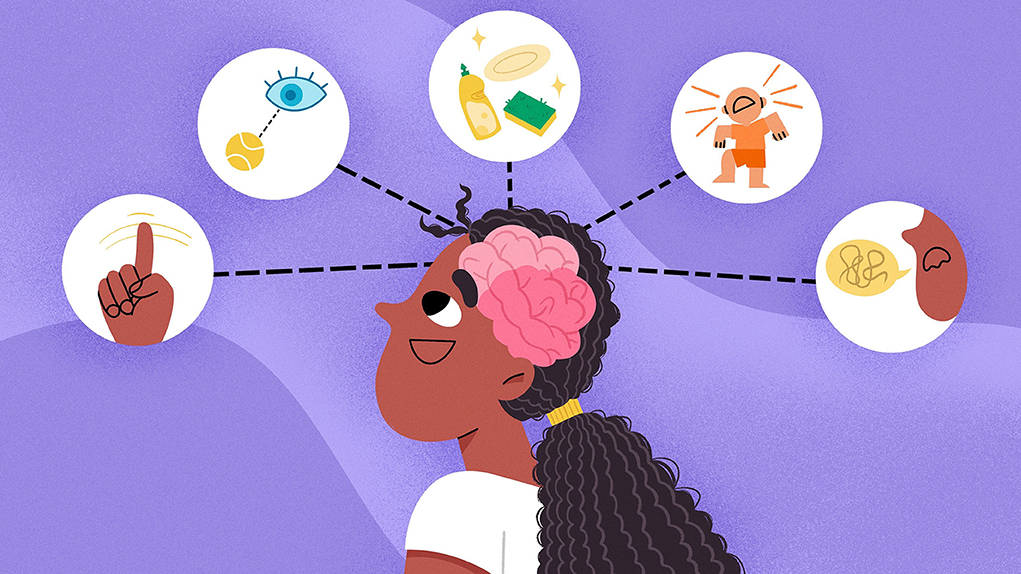
Every day, your brain accomplishes a lot. When your tummy growls, your brain registers this feeling as “time to eat”. When you feel super tired after coming home from school, your brain tells your body it is time to take a nap. Your brain also reminds you to complete tasks such as brushing your teeth, checking your Instagram DMs, and making sure you do the dishes before your parents get home. In a sense, you can picture your brain as the powerhouse of your body that drives your everyday activities. This article will focus on a section of the brain called the frontal lobe and how you may have used it, or seen others use theirs, daily.
The Frontal Lobe
The brain is responsible for many functions, and certain sections of the brain manage specific functions. There are a total of four brain sections, or lobes. These four sections are called the frontal, parietal, occipital, and temporal lobes (Figure 1A).
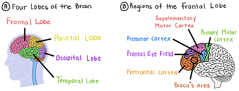
- Figure 1 - Breaking down the lobes of the brain.
- (A) The four lobes of the brain. (B) The five regions of the frontal lobe (Ballard, T., and Tucker, T. 25 Nov 2024. Image modified with Canva and Goodnotes).
Your frontal lobe is located approximately from the bottom of your forehead to the middle of the top of your head (Figure 1B). Your frontal lobe is responsible for motivation, planning, social behavior, and language/speech. Damage to the frontal lobe can lead to frontal lobe syndrome where your frontal lobe functions abnormally [1]. Symptoms include changes to your personality, loss of motivation to perform tasks, emotional outbursts, and depression.
Your frontal lobe is divided into five regions: the prefrontal cortex, the frontal eye field, the premotor cortex/supplementary motor cortex, the primary motor cortex, and The Broca’s area. The next few sections will discuss each of these five regions.
The Prefrontal Cortex
When your younger sibling or a child younger than you misbehaves, your immediate thought is to scold them. Sometimes, you have to tell them multiple times to stop whatever they are doing, until they understand what they are doing is not right. Sometimes they even throw a tantrum (Figure 2A). The reason younger children tend to take longer to understand what is right and what is wrong is because their prefrontal cortexes have not fully developed.
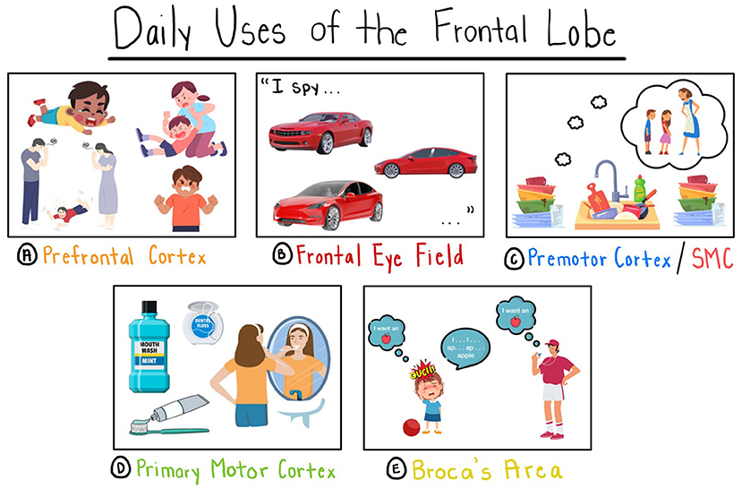
- Figure 2 - Daily life examples of the use of the frontal lobe.
- (A) Prefrontal cortex: children throw tantrums because their prefrontal cortexes are not fully developed. (B) Frontal eye field: playing the game “I Spy”. (C) Premotor cortex/SMC: planning to do the dishes before mom gets home. (D) Primary motor cortex: brushing your teeth. (E) Broca’s area: trying to say a phrase after a concussion (Ballard, T., and Tucker, T. 25 Nov 2024. Image modified with Canva and Goodnotes).
Your prefrontal cortex is located in the front part of your brain, where your forehead is. It is responsible for thoughts, actions, behavior, and emotions [2]. When you mature, you tend to behave well because you use your prefrontal cortex more, to think before you speak or act in a situation. Your prefrontal cortex continues to grow until around age 25.
Stress is when something negative within your environment (such as having too much homework) or something physical (such as scraping your knee) causes your body to send a signal to your brain. When too many stress signals are sent to the brain, especially for a long time, this can cause problems for our brain. For example, if you smoke at a young age (or before your prefrontal cortex fully develops) then your prefrontal cortex will become stressed and shrink. As a result, you may participate in more rule-breaking behavior and possibly become addicted to smoking [3].
The Frontal Eye Field (FEF)
Do you remember the game “I Spy”? It begins with someone saying, “I spy with my little eye…” and finishes with naming any object you would have to search for (Figure 2B). When you search for this object, you use your frontal eye field (FEF) to move your eyes in the necessary directions. Your FEF is located right behind your prefrontal cortex and is responsible for moving your eyes voluntarily (on purpose). For example, in baseball, the catcher scans the air to catch the ball.
If stress or damage were to occur to the FEF, then you would not be able to move your eyes on purpose. For example, if you were the catcher and took a hard blow to your eye from a baseball, your eye might begin moving slower than usual. After that, while playing baseball, you would have trouble tracking the ball in the air to catch it—which is dangerous!
The Premotor Cortex/Supplementary Motor Cortex (SMC)
Have you ever woken up in bed and just laid there because you did not feel like getting up? You sit there for a few minutes to an hour thinking to yourself, “I really need to get up and get ready or I will be late”. During this time, you are planning your movements in your brain.
The action of thinking about and planning your movements takes place in your premotor cortex and supplementary motor cortex (SMC). Both brain sections are responsible for planning and organizing movements (Figures 2C, D, 3A). Your premotor cortex/SMC is located behind the frontal eye field.
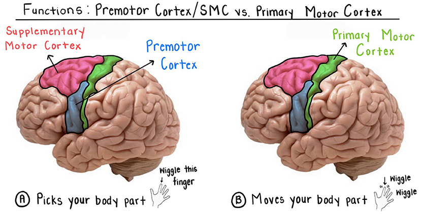
- Figure 3 - Functions of the premotor cortex/SMC and primary motor cortex.
- (A) Your premotor cortex/SMC chooses what body part it wants to move (for example, your pointer finger). (B) Your primary cortex moves the body part your premotor cortex/SMC area has chosen (your pointer finger wiggles) (Ballard, T., and Tucker, T. 25 Nov 2024. Image modified with Canva and Goodnotes).
Damage or stress to this brain area causes you to have trouble moving parts of your body [4]. Tourette’s Syndrome (TS) is a good example. TS is a disorder where your body moves or makes you say things without you meaning to. For example, a person who has TS may have random outbursts or turn their head repeatedly, even though they did not mean to. This happens because their premotor cortex/SMC constantly signals their body to perform these actions without their permission.
The Primary Motor Cortex
Imagine you are about to cross the finish line when running in a big race. You are coming close to finishing first, but you notice someone behind you who is trying hard to beat you. You know you will have to run harder to keep in first place, so you push yourself to run hard until you cross the finish line… coming in first!
The primary motor cortex is responsible for voluntarily moving your body parts. It is located in the back part of the frontal lobe, right behind the premotor cortex/SMC. As mentioned above, you use your premotor cortex/SMC to think about moving a certain body part. Afterwards, your premotor cortex/SMC sends a signal to your primary motor cortex to carry out the action (Figures 2C, D, 3B). For example, you use your premotor cortex/SMC to think about wiggling your pointer finger. Then your primary motor cortex wiggles your pointer finger (Figure 3).
Imagine you were a great piano player, but a tumor formed near your primary motor cortex. You sit at the piano bench attempting to play one of your favorite songs. Since the tumor formed, you have not been able to move your right hand as fast as you used to. When you try to play the song on the piano, you find yourself not being able to play the song correctly because you keep messing up with your right hand—which is frustrating!
The Broca’s Area (BA)
Try saying the word: “worcestershire”. You may find it difficult to say (The correct way to pronounce this word is: wor-stuh-sure). When trying to say this word, you use the muscles in your throat, tongue and mouth to pronounce the sounds of the word. The area within your frontal lobe responsible for speaking is your Broca’s area (BA).
The BA is located in the middle of your frontal lobe near the center of your brain. It is responsible for using your muscles to speak and for being able to speak. It is named after a French surgeon who discovered it in 1861, named Pierre Paul Broca [5].
Damage or stress to the BA will result in the inability to speak clearly. For example, say you were hit in the face by a dodgeball in gym class. Your teacher runs to you and tells you to say, “I want an apple”. You try to say this sentence but instead you jumble up your words (Figure 2E). It is evident that you have a concussion and need to see the nurse!
Putting It Together
Let us discuss one final time how you may have used or experienced each of these five brain regions on a daily basis (Figure 2). You might have seen the prefrontal cortex being used when a child throws a tantrum in the grocery store because they could not get the toy they wanted. When playing the game “I Spy”, you use your FEF to search for a red car, for example. When remembering to do the dishes before mom gets home, you use your premotor/SMC to plan this action. When brushing your teeth, you use your primary motor cortex to move your hand muscles. When trying to say the right words as you speak to people each day, you use your Broca’s area to think about what you are going to say and to say it.
Now that you have learned about the five regions of the frontal lobe, it is your turn to develop examples of how you may have used (or seen others use) each region daily. Ready, set, go!
Glossary
Frontal Lobe: ↑ Located behind your forehead and divided into five regions. Responsible for motivation, planning, social behavior, and language/speech.
The Prefrontal Cortex: ↑ Located in the front part of the brain where your forehead is. Responsible for thoughts, actions, behavior, and emotions.
The Frontal Eye Field: ↑ Located behind the prefrontal cortex. Responsible for moving the eyes voluntarily (on purpose) to look at objects and to pay attention to what is being shown.
The Premotor Cortex/Supplementary Motor Cortex: ↑ Located behind the frontal eye field. Responsible for planning and organizing movements using your visual field.
The Primary Motor Cortex: ↑ Located in the back part of the frontal lobe right behind the premotor cortex/SMC. Responsible for voluntarily moving your body parts.
The Broca’s Area: ↑ Located in the middle of your frontal lobe near the center of your brain. Responsible for using your muscles to speak and being able to speak.
Conflict of Interest
The authors declare that the research was conducted in the absence of any commercial or financial relationships that could be construed as a potential conflict of interest.
References
[1] ↑ Pirau, L., and Lui, F. 2023. “Frontal lobe syndrome”, in StatPearls (Treasure Island, FL: StatPearls Publishing).
[2] ↑ Arnsten, A. F. 2009. Stress signaling pathways that impair prefrontal cortex structure and function. Nat. Rev. Neurosci. 10:410–22. doi: 10.1038/nrn2648
[3] ↑ Xiang, S., Jia, T., Xie, C., Cheng, W., Chaarani, B., Banaschewski, T., et al. 2023. Association between vmPFC gray matter volume and smoking initiation in adolescents. Nat. Commun 14:4684. doi: 10.1038/s41467-023-40079-2
[4] ↑ Purves, D., Augustine, G. J., Fitzpatrick, D., Katz, L. C., LaMantia, A. –S., McNamara, J., et al. 2001. “Neuroscience. 2nd edition”, in The Premotor Cortex (Sunderland, MA: Sinauer Associates).
[5] ↑ Stinnett, T. J., Reddy, V., and Zabel, M. K. 2023. “Neuroanatomy, Broca Area”, in StatPearls (Treasure Island, FL: StatPearls Publishing).