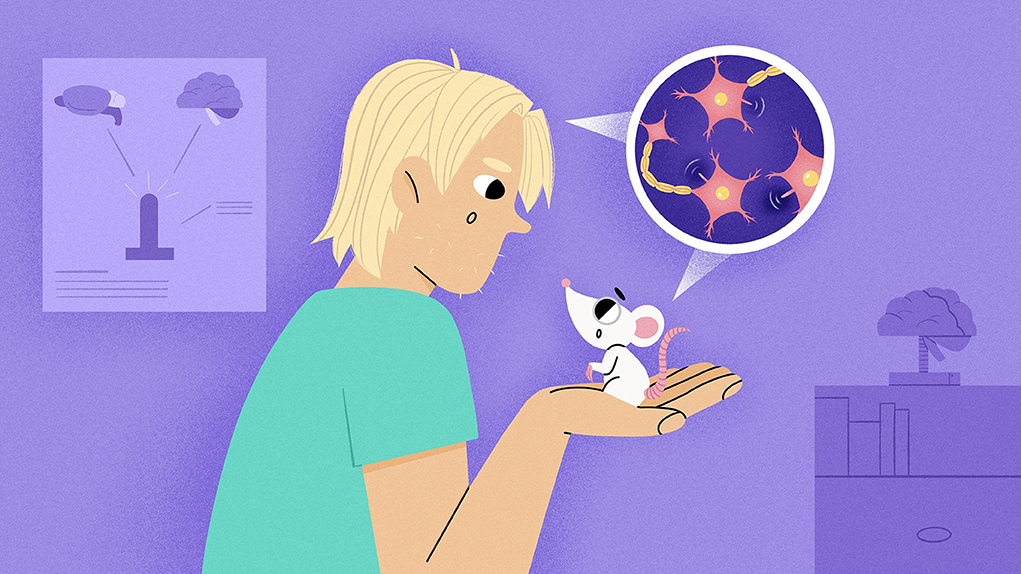
Abstract
Our brains are like incredibly complex puzzles, with billions of pieces that have been growing and developing since before we were born. But did you know that tiny, hair-like structures on our cells called primary cilia play a big part in this process? Primary cilia act as antennas, helping our brain cells communicate, travel, and even build connections, by guiding the assembly of this big puzzle. However, when primary cilia cannot form properly or cannot function smoothly, this can impact the development of many organs, including the brain. Scientists have found that shorter or fewer primary cilia are linked with conditions that may affect brain development, including a group of disorders named ciliopathies. By understanding the importance of primary cilia, we can find out more about brain development and the role cilia play in the assembly of this big puzzle.
How is the Brain Formed?
The human brain is truly remarkable, consisting of around 86 billion tiny cells that work together to control every function and action of our bodies. But how does such a complicated organ come to be? It all started before we were born, when we were just tiny embryos (Figure 1A). At this stage, stem cells are constantly dividing and creating all the various types of cells that make up our body parts. By receiving signals from surrounding cells, the stem cells in an embryo start to piece themselves together and become more specialized, taking on specific roles like transmitting information as neurons, supporting brain function as glial cells, or even forming muscles, skin, or blood. In the brain, the stem cells with some degree of specialization are called neural progenitor cells (NPCs) (Figure 1B).
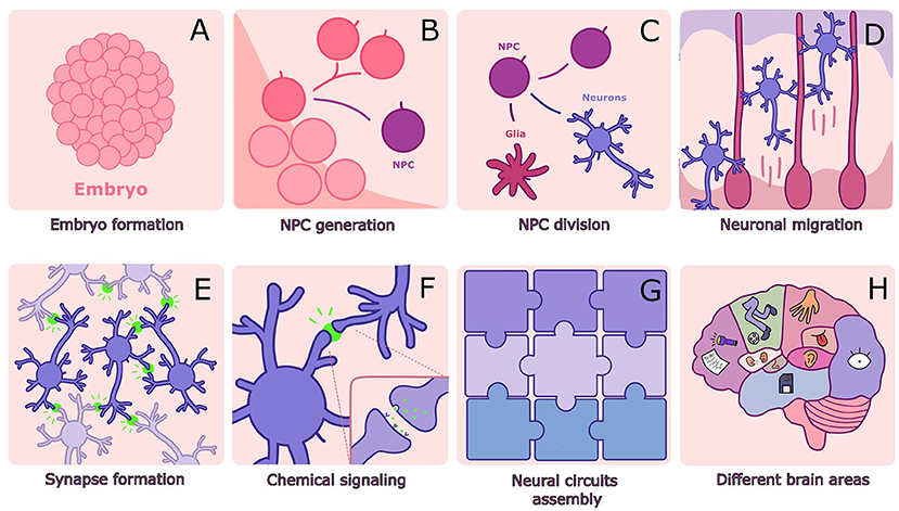
- Figure 1 - (A) The embryo is made of stem cells.
- (B) Stem cells in the embryo divide, and some become NPCs. (C) NPCs can divide to generate neurons and glial cells. (D) Newly formed neurons must migrate using glial cells as guides. (E, F) Neurons connect with other neurons through synapses, which allow communication using chemical signals. (G) Neurons connect like a large puzzle, where each piece is essential. (H) The completed puzzle forms the complete brain, with each region performing a specific function.
Neural progenitor cells have an extraordinary power: they can transform into two kinds of more complex cells—they can become either neurons or glial cells (Figure 1C). Neurons are specialized brain cells that can receive and send information, not only to other neurons but also to other types of cells. Neuron activity and communication allow us not only to think and create memories but also to see, move, talk, and even listen to sounds. Glial cells have other important roles, such as supporting neurons and cleaning up their waste products, protecting the brain from infections, helping fix things when there is a problem, and ensuring everything runs smoothly in the brain. In a sense, glial cells are like the glue keeping the neurons well attached—that is precisely what the word “glia” means (glue, in Greek).
When a new neuron is formed in a developing brain, it needs to travel to its specific place (Figure 1D). As neurons piece themselves together to form the structure of the brain, connections between neurons form, which are called synapses (Figure 1E). Synapses make it possible for neurons to communicate with each other, mostly through chemical signals (Figure 1F). Like puzzle pieces, neurons need to fit together effectively and build just the right number of connections. As more and more neurons connect with each other, a big, intertwined web is created, forming what are known as neural circuits (Figure 1G). When this happens, the different parts of the brain become organized into regions that may each play a more specific role in thinking, learning, speaking, seeing, or controlling body movements (Figure 1H).
However, brain development does not stop there. As we grow and learn new things and develop new interests like sports or music, our neurons can form even more synapses. This ability to change connections between neurons is called plasticity, and in this way the brain is always changing and adapting—thanks to the hard work of brain cells and their special “antennas”, the primary cilia.
What are Primary Cilia and How Do They Participate in Brain Formation?
Primary cilia are tiny, antenna-like structures, with each cell having just one. They help the cell sense signals and communicate, like mini satellite gathering information. For neurons and glia, this antenna helps send and receive signals between brain cells. This function is especially important when the brain is developing because primary cilia help cells capture important chemical messages that tell them when to perform certain tasks, such as dividing and making more copies of themselves, becoming more complex “adult” cells (which is called differentiation), or migrating to a specific place in the brain (Figure 2) [1–3].
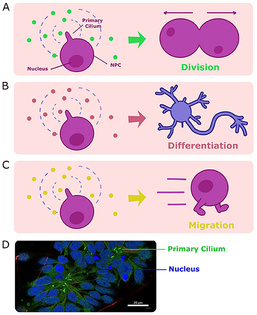
- Figure 2 - When primary cilia on NPCs detect certain signals in their environment, they tell the cell to either (A) divide, (B) differentiate into neuronal cells, or (C) migrate to a different location in the brain.
- All these processes are crucial for healthy brain formation. (D) A microscope image showing healthy cells, with primary cilia colored green and the nucleus colored blue. Each cilium measures about 5 micrometers in length—about 1/4 the diameter of a strand of hair (microscopic image collected by the Peça Lab at CNC-UC).
For example, the primary cilia of NPCs tell them when to divide into either more NPCs or into neurons or glia, which do not divide anymore. This way, the brain has a mechanism to ensure that it has precisely the right number of cells it needs to work in a healthy way. After neurons differentiate, they need to know where to go in the brain—that is where their antennas come into play once more. Primary cilia capture signals from neighboring cells that tell these neurons where they should go and even where to form new synapses. Based on the signals that primary cilia collect, more synapses are made, and these connections help to create mature neural circuits.
Overall, primary cilia play an important role in putting together and organizing the puzzle pieces that form the brain, and it is very important that they do this job just right. When a piece is out of place, out of order, or even when there are too many pieces, health problems can result.
What Happens When Primary Cilia Do Not Work Properly?
When primary cilia are either formed incorrectly or cannot be formed at all, they will not function properly, and diseases can result, including a group of diseases called ciliopathies. Ciliopathies can affect a few organs or multiple organs, including the brain, liver, and kidneys; in severe cases it can even be lethal for the developing embryo. When the brain is affected by ciliopathies, its puzzle pieces—the neurons and glia—have trouble connecting and communicating with each other. If this happens, the person will have difficulties with some activities, like learning new things. Altered primary cilia are also linked to neurodevelopmental disorders such as autism spectrum disorder and intellectual disability, in which brain development is impaired. Neurodevelopmental disorders can affect not only how people learn but also the ability to communicate or even move, making life more challenging.
To learn more about neurodevelopmental disorders and primary cilia, scientists did experiments using mouse brain cells and even human cells from individuals with disorders including fragile X syndrome, Rett syndrome and MBD5-associated neurodevelopmental disorder (MAND) [4–6]. These scientists made an intriguing discovery—the primary cilia in these cells appeared to be shorter and fewer in number compared to cells from healthy mice or people without these disorders (Figure 3). Since primary cilia play a crucial role in brain development, these findings suggest that primary cilia could be involved in these neurodevelopmental disorders, as the brain cells might become “lost” without functional antennas.
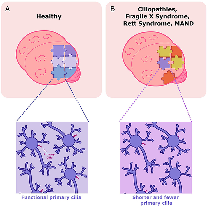
- Figure 3 - (A) In a healthy brain, each brain cell has a functional primary cilium and the brain cells come together properly, completing the “puzzle”.
- (B) In diseases like ciliopathies, fragile X syndrome, Rett syndrome, or MAND, neurons have fewer primary cilia or these structures can be shorter or even absent. In these cases, the brain’s puzzle pieces might not assemble correctly.
Scientists are working hard to understand more about these special cellular antennas that help healthy brains develop. We know now that they are crucial for neuron division, differentiation, and migration. However, we do not know exactly which chemical signals are involved in this communication. By studying more patients and coming up with creative ways to study this problem, scientists can generate new ideas for research, which could eventually lead to treatments for people with these conditions. Uncovering the secrets of the tiny primary cilia will bring us a step closer to solving the incredible and complex brain puzzle.
Glossary
Embryos: ↑ The earliest stage of development after fertilization in many species, where a group of cells has the potential to grow into a complete organism.
Stem Cells: ↑ Stem cells are cells that can become many different types of cells in the body. They play a key role in development, growth, and repairing damaged tissues or organs.
Neurons: ↑ Neurons are nerve cells that carry messages in the brain and throughout the body. They help us think, move, feel, and remember by using mostly chemical signals to communicate.
Glial Cells: ↑ Glial cells are important support cells in the brain. They protect neurons, provide them with nutrients, clean up waste, and help them communicate, ensuring the brain stays healthy and works well.
Neural Progenitor Cells (NPCs): ↑ Cells in the developing brain that can either divide or generate brain cells called neurons and glial cells.
Synapses: ↑ A small gap between neurons where signals are transmitted, enabling brain cells to communicate and coordinate functions such as thinking, moving, and sensing.
Neural Circuits: ↑ Neural circuits are networks of connected brain cells that send and process information, like tiny electrical highways. They control everything from breathing to thinking, helping the brain respond to the world.
Differentiation: ↑ The process where stem cells become specialized, turning into different types of cells with specific jobs, like neurons or glial cells.
Ciliopathies: ↑ A group of rare disorders where the primary cilia do not work properly, causing various problems.
Neurodevelopmental Disorders: ↑ Conditions that affect brain growth and function, often starting in childhood. They can impact learning, behavior, or movement, like autism spectrum disorder and intellectual disability.
Primary Cilium: ↑ A tiny antenna-like structure on each cell that helps receive signals and communicate. The plural is primary cilia.
Conflict of Interest
The authors declare that the research was conducted in the absence of any commercial or financial relationships that could be construed as a potential conflict of interest.
References
[1] ↑ Adkins, G. J. J., Doherty, D., and Hevner, R. F. 2012. Joubert syndrome: brain and spinal cord malformations in genotyped cases and implications for neurodevelopmental functions of primary cilia. Brain Struct. Funct. 217:695–709. doi: 10.1007/s00401-012-0951-2
[2] ↑ Hasenpusch-Theil, K., and Theil, T. 2021. The multifaceted roles of primary cilia in the development of the cerebral cortex. Front. Cell Dev. Biol. 9:1–14. doi: 10.3389/fcell.2021.630161
[3] ↑ Higginbotham, H., Eom, T. Y., Mariani, L. E., Bachleda, A., Hirt, J., Gukassyan, V., et al. 2012. Arl13b in primary cilia regulates the migration and placement of interneurons in the developing cerebral cortex. Dev. Cell. 23:925–38. doi: 10.1016/j.devcel.2012.09.019
[4] ↑ Lee, B., Panda, S., and Lee, H. Y. 2020. Primary ciliary deficits in the dentate gyrus of Fragile X Syndrome. Stem Cell Reports. 15:454–66. doi: 10.1016/j.stemcr.2020.07.001
[5] ↑ Frasca, A., Spiombi, E., Palmieri, M., Albizzati, E., Valente, M. M., Bergo, A., et al. 2020. MECP2 mutations affect ciliogenesis: a novel perspective for Rett syndrome and related disorders. Front. Cell Dev. Biol. 8:1–18. doi: 10.15252/emmm.201910270
[6] ↑ Martins, M., Oliveira, A. R., Martins, S., Vieira, J. P., Perdigão, P., Fernandes, A. R., et al. 2023. A novel genetic variant in MBD5 associated with severe epilepsy and intellectual disability: potential implications on neural primary cilia. Int. J. Mol. Sci. 24:12603. doi: 10.3390/ijms241612603