
Abstract
Your brain can be divided into various areas, one of which is responsible for your sense of touch. This part of your brain can be divided into even smaller areas that communicate with each body part. We can use a special map of the human body, called a sensory homunculus, to help us understand the various sizes of these parts of the brain. We will explain how this map was created and tell you about research showing how these brain areas can change. One study showed that brain areas can be recycled, meaning that the brain areas that no longer receive messages from the body can be used by other functioning brain areas. Another study showed that these changes can even occur within a single day! These studies can help scientists to better understand the brain and to help people who have problems with the sense of touch.
Your Brain–An Overview
Your brain weighs about three pounds and feels like Jell-O. Unlike Jell-O, your brain can complete many tasks. The outer layer of the brain is called the cortex. The cortex is made of 80 billion nerve cells called neurons. Groups of neurons in your brain send messages to and from other neurons located all over your body. Some of the neurons outside of your brain contain sensors that respond to pressure on your skin. When activated, these neurons send electrical messages back to your brain to let you know you have touched something (or that something has touched you). The area of the brain responsible for interpreting your sense of touch is called the sensory cortex. The sensory cortex is divided into specific areas, each of which communicates with a single part of your body, such as your left pinky finger or your tongue.
Early Attempts to Divide the Brain
In 1909, Dr. Korbinian Brodmann was the first person to discover that the cortex is divided into specific areas based on the shape of neurons [1]. Dr. Brodmann divided the brain into 52 areas and the sensory cortex contains three of these areas (Figures 1A,B). You have a sensory cortex on both sides of your brain (Figures 1B,C). Dr. Brodmann’s discovery is still important today. He showed that the brain is filled with different types of neurons that are arranged in groups. The existence of these groups of neurons led scientists to believe that different areas of the brain are responsible for different body functions.
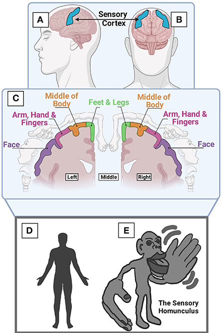
- Figure 1 - (A) Side view and (B) front view of the brain, showing the sensory cortex.
- (C) Brain slice showing how the sensory brain areas are arranged. Notice that this arrangement is similar to the way body parts are arranged. (D) A normal-sized human. (E) The sensory homunculus, which is a kind of map showing what a human would look like if the size of body parts was based on the number of neurons in that brain area. Larger size means the body part both has more neurons in its brain area and has a better sense of touch. (Created with BioRender.com).
Dr. Wilder Penfield was a brain doctor who accidentally helped us understand the function of the sensory cortex. In the 1950s, Dr. Penfield helped patients who had a brain disease that caused irregular movements. He determined which brain area was sending “bad” messages to the body, by applying a small amount of electricity to several areas of his patients’ brains. Curiously, he discovered that applying electricity to a specific part of the sensory cortex caused patients to feel a sensation in a specific body part [2]. He performed this on many sensory brain areas. As a result, he created a map showing which areas of the sensory cortex are responsible for the sense of touch in each limb (Figure 1C). Without Dr. Penfield’s map, we would not know that the brain is organized into specific areas that communicate with various parts of the body.
Your Brain’s Sensory Areas and A Strange-Looking Human
The organization of the sensory cortex reflects on the placement of each limb of your body (Figure 1C). For example, the “feet” area is far away from the “head” area. We can divide the sensory cortex areas even further. For example, each of your fingers has a unique area in the sensory cortex, and these areas are arranged in the brain similarly to the way they are arranged on your hand.
If the size of our body parts was based on the number of neurons in its brain area, instead of looking like we normally do (Figure 1D), we would like Dr. Penfield’s sensory homunculus (Figure 1E). In this model, the more neurons a sensory area has, the larger the body part is drawn. A larger size on this figure also means that the body part is more sensitive to touch.
At-Home Experiment
The size of brain areas associated with various body parts can be tested with an experiment that you can do at home. Have an adult unravel a paper clip so that there are two pointed ends that are a quarter inch apart from each other. Close your eyes and let the person randomly (and carefully!) poke you with either one or two points. See if you can sense whether one or two points of the paperclip are touching you. Repeat this process a few more times and keep track of whether you are wrong or right. Try this experiment on a sensitive area first (like a finger) and then on a less sensitive area (the upper arm). You might notice that your accuracy is much better for the fingers. This is because they have more neurons in their brain areas.
Use it or Recycle it
When part of the sensory cortex is not used for a long time, the area changes. Instead of “throwing out” the area of the brain that is no longer used, the brain “recycles” the neurons. In one experiment, researchers studied the brains of monkeys that had lost their middle fingers [3]. These monkeys were treated ethically and were still able to perform daily activities such as walking, swinging, eating, and playing with their monkey friends.
The scientists used a technique similar to Dr. Penfield’s to determine the size of each finger area in the brain. Instead of applying electricity directly to the brain, they applied a small, safe amount of electricity directly to the monkeys’ fingertips. They could determine the location in the brain where the electric signal traveled to. They repeated this process until they had enough points on each finger to create a map of the brain’s finger areas (Figures 2A,B).
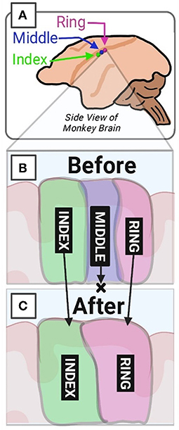
- Figure 2 - The brain “recycles” areas that are not used.
- (A) Side view of a monkey brain showing the sensory areas for the ring, middle, and index fingers. (B) The normal size of three finger areas in the brain. (C) The size of the index and ring finger areas in monkeys with no middle fingers. Notice how the sizes of the index and ring finger areas grew and they are now touching. (Created with BioRender.com).
The researchers discovered that, without touch information from the middle finger, the middle finger area of the brain shrunk. The area of the brain that represented the index and ring fingers grew, by “recycling” the neurons from the missing middle finger (Figure 2C).
Fast and Reversible Changes
The same kinds of brain changes occur in humans. In humans, these changes are measured using a technique called functional magnetic resonance imaging (fMRI) (Figure 3A) [4]. This technique can be used to look at brain activity in real time, so it is a useful tool for scientists who want to study changes in the brain without opening the skull.
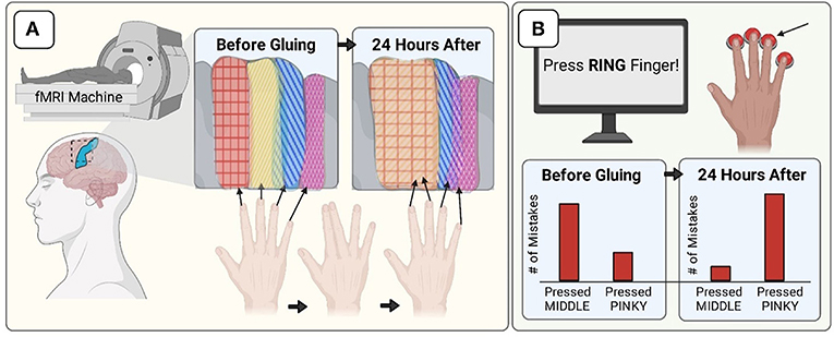
- Figure 3 - Gluing fingers together “glues” those finger areas of the brain together.
- (A) Before gluing, the middle and ring finger areas overlap. Twenty-four hours later, the brain areas of the index and middle fingers overlap, and the ring and pinky areas also overlap. (B) In the game, the computer indicated which finger to press. Before gluing, subjects more often accidentally pressed their middle fingers when asked to press their ring fingers. Twenty-four hours later, subjects more often accidentally pressed their pinkies. (Created with BioRender.com).
In one human experiment, researchers investigated the finger areas of the sensory cortex. Using fMRI, they determined the location and size of each brain area based on movement of the subjects’ fingers. The goal of the study was to determine whether humans could change the size of each brain region in 1 day.
Try this: First, lay your hand flat and palm-side-down on a table. Try moving your middle finger up and down while keeping the rest of your fingers flat on the table. Is this difficult? Next, try moving your ring finger up and down while keeping the rest of your fingers still. Was this easier or harder? You will probably find that moving your middle finger without moving your other fingers was easy, but moving your ring finger without moving your middle finger or pinky finger is more difficult. This behavior is also represented in the brain: the middle finger area and ring finger area overlap, which makes it more difficult for you to move these fingers separately.
To cause changes in the brain, the researchers glued the right index and middle fingers together. They used fMRI before and after the gluing period to determine the sizes of the finger areas. They found that, after the fingers had been glued for 24 h (and then unglued), the areas of the two previously-glued fingers (index, middle) now overlapped (Figure 3A).
Additionally, the researchers had the same people play a game before and after the 24-h gluing time. The subjects’ fingers were separated before playing the game the second time. Subjects had to press the button under the finger that was indicated on the computer screen, in less than 1 s (Figure 3B). This process was repeated many times.
The most interesting results came from the times when subjects needed to press their ring fingers. Before gluing, the subjects would accidentally press their middle fingers when they meant to press their ring fingers. This makes sense because the areas overlap in the brain. However, after the 24-h gluing period, the subjects accidentally pressed their pinky fingers (more than their middle fingers) when trying to press their ring fingers. This makes sense because the pinky and ring areas now overlap. Overall, these researchers demonstrated that changes in brain areas can cause changes in behavior and can occur over a single day!
Why Does This Matter?
The sense of touch is something we often take for granted. Without your sense of touch, you would not be able to sit up properly, and you would not be able to do something as simple as holding a cup of water without looking at it directly.
Some people are unable to interact with their environments the way others can because they have lost their sense of touch or possibly have lost a limb. Understanding how our brains communicate with our limbs to create the sense of touch allows scientists to create electronic devices that imitate the sense of touch. More recent research has used the ideas outlined in this paper to retrain the brain, to help patients who have lost the sense of touch to recover it. This work is promising and exciting, but there is still a lot we do not know. We need the unique “touch” of young minds like yours to help solve the questions researchers still face as they continue studying the sense of touch.
Glossary
Cortex: ↑ The outermost layer of the brain that is responsible for processing and sending messages about high-level processes such as decision-making, movement, emotion, and sense of touch.
Neurons: ↑ Cell that makes up the brain and communicates between the brain and the limbs of the body using electrical messages.
Sensory Cortex: ↑ The part of the brain that translates messages about pressure from the body; it can be divided into areas that get messages from specific body parts.
Sensory Homunculus: ↑ A model that shows what we would look like if the size of each body part was proportionate to the number of neurons in its corresponding brain region.
Functional Magnetic Resonance Imaging (fMRI): ↑ A technique that brain researchers use to observe brain activity in humans while it is happening.
Conflict of Interest
The authors declare that the research was conducted in the absence of any commercial or financial relationships that could be construed as a potential conflict of interest.
References
[1] ↑ Brodmann, K., and Gary, L. J. 2006. Brodmann’s Localisation in the Cerebral Cortex: The Principles of Comparative Localisation in the Cerebral Cortex Based on Cytoarchitectonics. Berlin; Heidelberg: Springer.
[2] ↑ Wilder, P., and Jasper, H. 1954. Epilepsy and the functional anatomy of the human brain. AMA Arch. Neurol. Psychiatry. 72:663–4. doi: 10.1001/archneurpsyc.1954.02330050133021
[3] ↑ Merzenich, M. M., Nelson, R. J., Stryker, M. P., Cynader, M. S., Schoppmann, A., and Zook, J. M. 1984. Somatosensory cortical map changes following digit amputation in adult monkeys. J. Comp. Neurol. 224:591–605. doi: 10.1002/cne.902240408
[4] ↑ Kolasinski, J., Makin, T. R., Logan, J. P., Jbabdi, S., Clare, S., Stagg, C., et al. 2016. Perceptually relevant remapping of human somatotopy in 24 hours. eLife. 5:e17280. doi: 10.7554/eLife.17280