
Abstract
Tooth enamel is the hardest substance in the human body. It begins forming during pregnancy and the way it forms can provide valuable information about early life. The cells that produce enamel are extremely sensitive to disturbances, so when a child experiences some kinds of stress during enamel development (childhood diseases, fever, or low concentration of oxygen in the blood for example), layers of enamel may contain “scars” reflecting the stressful experience. The type of enamel scars can tell dentists a story about what may have happened before the person was born or shortly after birth. Understanding enamel development is important for knowing the difference between normal enamel and abnormal changes, and such knowledge can help dentists to diagnose and treat enamel issues, to keep the teeth healthy.
Formation of Teeth
Children’s teeth are a valuable source of information about early development, especially when combined with the health records of the baby and the mother [1]. Formation of baby teeth and some permanent teeth begins during pregnancy and takes place in several stages. When certain unfavorable conditions arise during pregnancy, birth, or early childhood, tooth development can change. Studying such changes can give dentists and scientists clues about the conditions of a child’s early life!
Teeth are made of several materials. Dental pulp, which is the inner part of the tooth, contains blood vessels and nerves responsible for sensations such as pain, cold, and heat. Dentin is a mineralized substance around the pulp that protects the soft tissue. Enamel, which is considered the hardest substance in the human body, covers the surface of the tooth exposed to the mouth and protects the tooth’s deeper tissues (Figure 1) [2].
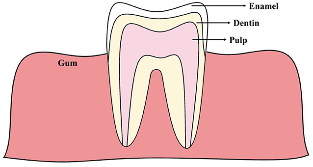
- Figure 1 - The structure of a tooth.
- Enamel is the structure that covers the visible surface of the tooth (which is also called the crown), and it is the hardest substance in the human body. Dentin is the second layer of the tooth, underneath the enamel, that protects the pulp tissue. The pulp is the inner part of the tooth, and it is made of cells, blood vessels, and nerves.
Because tooth enamel is such a tough substance, it remains preserved over time and can provide a lot of scientific information about early human development [3].
How is Enamel Formed?
In the early stages of development before birth, the human embryo is formed by three original layers of tissue that eventually develop into all the body’s organs and structures. The outermost tissue layer of the embryo is responsible for the formation of body parts including the brain, hair, nails, and dental enamel.
Ameloblasts are the cells responsible for the production of dental enamel. The process of enamel formation can be divided into three phases. The first phase is called the secretory phase, during which ameloblasts release proteins that make up the “foundation” of the enamel. At this stage, the tooth is formed but it has not yet hardened—it has a jelly-like consistency. For teeth to harden, a process called biomineralization must occur in the second phase of enamel production. During biomineralization, enamel acquires minerals like calcium and phosphate, which transform the tooth into a solid structure. Finally, during the maturation phase, the enamel undergoes further biomineralization, incorporating even more minerals into its structure.
Once teeth emerge in the mouth, the ameloblasts vanish. This means that, once the enamel is formed, it cannot be regrown. Enamel formation is sensitive to many conditions, including things that are happening in the body or in the environment while ameloblasts are making enamel. Thus, if unusual conditions occur while ameloblasts are active, it can affect enamel formation. This is why the enamel can act as an indicator of unhealthy events or conditions that occurred during a specific period in a person’s life [2].
The Neonatal Line
Ameloblasts create enamel in a cycle, alternating between periods of work and rest. This cycle causes the enamel to form in layers, which can be seen as a series of lines called growth lines. When problems occur during enamel formation, these growth lines may appear wider or darker, and they are then called stress lines. The neonatal line is one of the most prominent stress lines, found frequently in baby teeth and to a lesser extent in permanent first molars (Figure 2) [4]. The neonatal line forms during the stressful period experienced by the baby when it is being born. This means that the neonatal line signifies the transition between enamel formed during pregnancy and enamel that develops after the child’s birth [1].
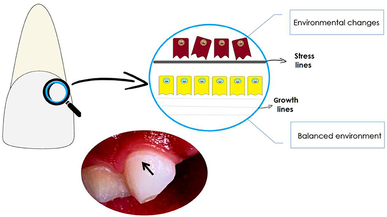
- Figure 2 - Ameloblasts lay down enamel in layers called growth lines.
- Because ameloblasts are highly sensitive cells, certain changes in the environment can cause problems during enamel formation. Consequently, the layers of enamel can become thicker or darker, resulting in the formation of stress lines. One of the most prominent stress lines is the neonatal line, which reflects the stress of birth. In the photo, the black arrow indicates the neonatal line on a baby’s tooth.
Studies show that children whose mothers suffered from depression or anxiety during pregnancy, or children who had mothers with a history of severe depression or serious mental health issues, had wider neonatal lines than children whose mothers did not have these conditions. Narrower neonatal lines were also found to be strongly associated with high levels of family social support, which is believed to be protective against depression. Therefore, the neonatal line can be considered a bodily indicator of a disturbed environment around the time of birth [4].
Enamel Defects
Disturbances during enamel production can lead to tooth scars that can tell dentists about the conditions a person experienced during childhood. When enamel defects are limited to a single permanent tooth, a local factor may have caused the defect, such as an injury to that tooth or an infection in the area where the tooth was developing. When multiple teeth have enamel defects, this likely means that body-wide issues occurred in the early years of life when the teeth were developing. These situations could include lack of oxygen, premature birth, use of certain medications, severe fever, serious infections, nutritional deficiencies, or even mutations in certain genes [5, 6].
If any of these factors occur in the secretion stage of enamel production, they can cause an enamel defect called hypoplasia, in which not enough enamel is produced. On the other hand, when negative factors are experienced during the mineralization stage, this can lead to hypomineralization, in which the enamel that forms is of poor quality, with fewer minerals incorporated [5]. Basically, enamel defects that happen during development look like scars on the teeth, telling dentists that, at some point, there were changes that affected enamel production (Figure 3).
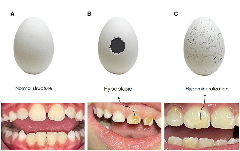
- Figure 3 - Disturbances in enamel production can lead to tooth defects.
- If we compare tooth enamel to an eggshell, there are three possible situations. (A) The enamel structure is intact, like an undamaged eggshell. (B) There is a gap in the enamel, indicating a defect in the amount of enamel made. This condition is called hypoplasia. (C) The enamel is present, but low quality and fragile, like a very thin eggshell. This happens when not enough minerals are added to the enamel, and it is called hypomineralization.
Why is it Important to Know About Enamel Formation?
Tooth formation, especially the formation of enamel, is a complex process that is sensitive to environmental changes. This means that changes in the normal structure of tooth enamel can provide valuable information about events or conditions that occurred both before birth and during early childhood. Understanding the processes involved in enamel development is important for recognizing the difference between normal tooth development and unhealthy changes. Today, enamel defects affect many children. Early diagnosis of unhealthy enamel changes can help reduce the tooth damage caused by these defects and can help with the development of new approaches to treat enamel disorders—keeping kids’ teeth healthy!
Glossary
Enamel: ↑ Mineralized hard structure that covers the visible part of the teeth.
Ameloblasts: ↑ Cells that create the enamel layer of the teeth.
Biomineralization: ↑ Process by which minerals such as calcium and phosphate are added to the enamel, to make it an extremely hard substance.
Growth Lines: ↑ Lines that can be seen in tooth enamel using a microscope.
Stress Lines: ↑ Growth lines that are thicker than normal and can be seen with the naked eye.
Neonatal Line: ↑ A common type of stress line in baby teeth that indicates the stress of being born.
Hypoplasia: ↑ An enamel defect in which not enough enamel is created.
Hypomineralization: ↑ An enamel defect in which not enough minerals are incorporated into the enamel, so that the enamel that exists is of poor quality.
Conflict of Interest
The authors declare that the research was conducted in the absence of any commercial or financial relationships that could be construed as a potential conflict of interest.
The author(s) declared that they were an editorial board member of Frontiers, at the time of submission. This had no impact on the peer review process and the final decision.
Acknowledgments
Financial support from FAPESP (2023/12014-8), CNPq (PIBIC-EM), and CAPES Foundation.
References
[1] ↑ Behie, A. M., and Miszkiewicz, J. J. 2019. Enamel neonatal line thickness in deciduous teeth of Australian children from known maternal health and pregnancy conditions. Early Hum. Dev. 137:10482. doi: 10.1016/j.earlhumdev.2019.07.004
[2] ↑ Nanci, A. 2018. “Enamel: composition, formation, and structure”, in Ten Cate’s Oral Histology. 9 th ed. (St. Louis, MO: Elsevier) p. 118.
[3] ↑ Porto, I. M., Laure, H. J., Tykot, R. H., de Sousa, F. B., Rosa, J. C., Gerlach, R. F. 2011. Recovery and identification of mature enamel proteins in ancient teeth. Eur. J. Oral. 1:83–87. doi: 10.1111/j.1600-0722.2011.00885.x
[4] ↑ Mountain, R. V., Zhu, Y., Pickett, O. R., Lussier, A. A., Goldstein, J. M., Roffman, J. L., et al. 2021. Association of maternal stress and social support during pregnancy with growth marks in children’s primary tooth enamel. JAMA Netw. Open. 11:e2129129. doi: 10.1001/jamanetworkopen.2021.29129
[5] ↑ Patel, A.; Aghababaie, S.; Parekh, S. 2019. Hypomineralisation or hypoplasia? Br. Dental J. 227:683–686. doi: 10.1038/s41415-019-0782-9
[6] ↑ Gonçalves, J. L., Duarte, A. C. A., Almeida-Junior, L. A., de Carvalho, F. K., de Queiroz, A. M., Arnez, M. F. M., et al. (2022). Enamel biomineralization under the effects of indomethacin and celecoxib non-steroidal anti-inflammatory drugs. Sci. rep. 12:15823. doi: 10.1038/s41598-022-19583-w