Abstract
Ultrasound machines use sound waves to take a look inside your body. These sound waves are very high pitched and cannot be heard or felt. They are generated by an ultrasound machine that uses crystals at the end of an ultrasound probe. These waves pass through your skin and as they reflect back to the machine from tissues in your body, the machine turns them into an image. In this article, you will learn about the different uses of ultrasound, like seeing babies inside the uterus or examining important body organs in patients who are feeling sick. You will also learn about the safety of ultrasound in case you ever need to get one. Last, we will explore the future of ultrasound in medicine by learning about point-of-care ultrasound, or POCUS, which is making it easier for children and grownups to get ultrasounds when they need them!
What is Ultrasound?
Sound is everywhere around us. We can hear some of the sounds around us, but many sounds are completely undetectable. When sound is made by animals or objects (for example, a barking dog or the sirens of an ambulance), it passes through the air in the form of invisible waves. These sound waves travel until they are heard by a receiver, like your ear. Scientists can study sounds by measuring the size and energy of the waves that are created. Just like your height and weight can be measured by your doctor, a sound wave’s length and frequency can be measured by scientists. Wave frequency refers to the length of a wave and how fast it is traveling. Sound wave frequency is measured using a unit called Hertz (Hz). Our ears can hear audible sound, which are sounds with frequencies that range between 20 and 20,000 Hz. Any sound frequencies above or below that cannot be heard by our ears. Sound waves with frequencies lower than 20 Hz are called infrasound and sounds with frequencies >20,000 Hz are called ultrasound. Look at Figure 1 to learn about the different sound frequencies that were discussed here.
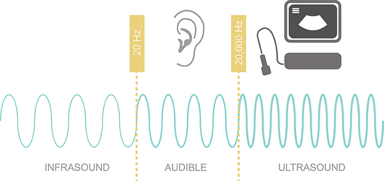
- Figure 1 - Different sound frequencies: infrasound has sound waves with frequencies lower than 20 Hz; audible sound, which has sound waves with frequencies between 20 and 20,000 Hz; and ultrasound, which has sound waves with frequencies >20,000 Hz.
If you stop reading this and let out a high-pitched scream, you can probably generate sound waves with frequencies of about 3,000 Hz. We can use special machines to generate ultrasound frequencies that are much greater than that. The machines we use to generate ultrasounds can create sound waves with frequencies above 1,000,000 Hz. These very high frequency sounds are especially useful in medicine because they can be used to safely look inside our bodies. Because sound has the ability to travel through air, liquids, and solids, we can point sound waves toward the heart, for example, and see what the naked eye cannot see through the skin.
How Does an Ultrasound Machine Work to Look Inside Our Bodies?
Imagine yourself standing in a valley between two large mountains. If you scream loudly, you will hear an echo of your voice. This is because the sound waves bounce back and forth between the mountains and return back to your ears. Similarly, an ultrasound machine produces sound waves and listens for the returning waves made by the sound bouncing back off the tissues that make up the different organs in your body.
In Figure 2, you can see an ultrasound probe that produces ultrasound waves. When it is pointed at your body, the sound waves can travel through your skin and into the organ that your doctor wants to look at (for example, your liver). You can think of the ultrasound probe as a “magic wand,” sending many ultrasound waves into your body to look at what is inside. Every time the waves hit a surface with different physical properties, they get reflected back to the probe [1]. The probe measures the amount of time it takes for each wave to return to it. Using this information, a computer is able to calculate the distance to each surface the wave bounces into. The computer puts all this information together and creates an image of what is deep inside your body.
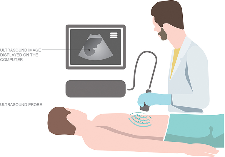
- Figure 2 - A physician uses an ultrasound machine by pointing the sound signal into the patient’s body.
- In this picture, the physician is looking at the patient’s liver!
How Can We Tell the Difference Between Tissues in the Body?
Different tissues in your body appear differently on an ultrasound machine. This is because the amount of sound that is reflected back from the tissue and heard by the machine depends on the type of tissue that the sound waves are hitting and the speed of the waves coming back to the probe [1]. The fat, air, and water content of the tissue determines that tissue’s density, which in turn affects the time it takes for ultrasound to be reflected back into the machine [2].
What is Ultrasound Used For?
In medicine, ultrasound waves can be used to identify why someone is sick—this is referred to as a diagnostic use. Ultrasound can also be used to treat someone who is sick—this is called therapeutic use [3].
Diagnostic Uses: Identifying Why Someone Is Sick
Ultrasound can be used by people in healthcare to understand why someone is sick. There are many body parts that can be looked at, including the heart, lungs, liver, gallbladder, kidneys, appendix, and even your muscles and joints!
For example, imagine you are having stomach pain. You go to the hospital and see a doctor to find out why your stomach is hurting. Your doctor may use an ultrasound machine to look into your abdominal region to see what may be causing you pain. Could it be that the pain is coming from your appendix? The appendix is a small part of your intestine and it can get infected. This is called appendicitis, and it can be very painful. To find out if you have appendicitis, a doctor might use an ultrasound machine to look for signs of appendicitis inside your abdomen. Some of these signs include thickening of the appendix, dilation (or widening) of the appendix, and fluid collection around the appendix. Looking for these signs using an ultrasound machine can help your doctor identify what is wrong and decide how to treat the problem to help you feel better.
Therapeutic Uses: Treating Sick People
Ultrasound can be used to treat illness. Ultrasound waves carry energy with them that can be focused on specific tissue. Ultrasound waves can be produced at very high frequencies (such as 4,000,000 Hz) to send energy into the body’s tissues. For example, sometimes little stones are formed when your kidneys are working to make urine. These kidney stones may not cause any problems for some people, but for others they can get stuck and block the flow of urine to your bladder, causing a lot of pain. Specialists can use high-energy ultrasound to break up the stones into smaller pieces that can be passed through in your urine with much less pain.
Ultrasound can also be used to guide treatment when doctors need to look inside the body to help treat an illness. For example, sometimes illnesses can cause fluid to collect in the abdomen and cause a lot of pain. When this happens, one way to help patients feel better is to drain this fluid. Ultrasound can be used to help the doctor look inside the body to find where the fluid is so that they can drain it at the appropriate site.
Is Ultrasound Safe?
Yes, ultrasound is very safe! Remember that ultrasound machines use sound waves. These sound waves have virtually no harmful effects on your body. The high-energy sound waves used by ultrasound machines might cause small amounts of heat to enter the body [3]. But thankfully this is not something to worry about, especially when an ultrasound is done safely by a trained professional.
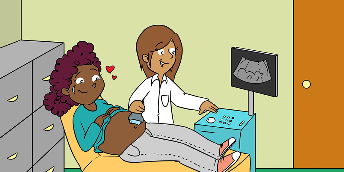
Ultrasound is very different from other ways that healthcare professionals look inside the body. Have you ever seen an X-ray? Just like ultrasound, X-rays provide a picture of what is going on in your body. While ultrasound uses sound waves, X-rays use light waves to look under your skin. Although we cannot see these light waves, they carry radiation energy, which can be harmful in high amounts. Ultrasound, on the other hand, is so safe that it is even used to look inside women who are pregnant and examine how their babies are developing!
We know that getting an ultrasound is safe—but does it hurt? Well, most ultrasound is done using a type of jelly that helps the ultrasound wand work better. This jelly feels like cold, wet goo, and it will touch your skin. But besides feeling the wand and the jelly, the sound waves passing from the ultrasound machine through your skin are completely painless, and you cannot even feel them.
What is the Future of Ultrasound?
Ultrasound is not new in medicine. We have actually been using ultrasound to take pictures of the inside of the body for over 50 years! But this does not mean that the technology inside ultrasound machines has stayed the same. Over time, as computers have gotten better, ultrasound machines have been able to take better quality pictures and videos of our bodies. The machines themselves have also gotten a lot smaller. Remember seeing pictures of the big, bulky cellular phones your parents had years ago? Now compare that with smart phones today—today’s phones are much smaller and faster! The same thing has happened with ultrasound machines! Smaller ultrasound machines mean more healthcare providers will be able to use them in different settings.
The use of smaller, more portable ultrasound machines is sometimes referred to as point-of-care ultrasound, or POCUS. This is where the real magic of ultrasound happens. Imagine going to see your doctor and finding out that you need an ultrasound test because you are having really bad stomach pain. Without POCUS, your doctor would have to send you somewhere else to have the test done. Depending on where you live, the closest place with an ultrasound machine might be a hospital far away. With POCUS, your doctor will be able to take a quick look inside your stomach with an ultrasound, right there in the office. This will allow your doctor to find out what is going on inside your body and start treating you a lot quicker. Because of these advantages, POCUS is already being used on people who are sick or injured, in emergency rooms and medical clinics all around the world. Many lives have been saved—and in the future, many more will be saved—thanks to the magic of sound waves!
Author Contributions
All authors contributed equally to this work.
Glossary
Frequency: ↑ A unit of measurement that is related to the length of a sound wave and how fast it is traveling. Sound wave frequency is measures using a unit called Hertz (Hz).
Audible Sound: ↑ Sound waves with frequencies between 20 and 20,000 Hz.
Infrasound: ↑ Sound waves with frequencies <20 Hz.
Ultrasound: ↑ Sound waves with frequencies above 20,000 Hz.
Diagnostic Use: ↑ Using a tool—in this article, ultrasound—to help identify why someone is sick.
Therapeutic Use: ↑ Using a tool—in this article, ultrasound—to treat people who are sick.
POCUS: ↑ Point-of-care ultrasound is the use of a smaller, more portable ultrasound machine at the bedside of a patient.
Conflict of Interest
The authors declare that the research was conducted in the absence of any commercial or financial relationships that could be construed as a potential conflict of interest.
Acknowledgments
We would like to acknowledge Ryan Giuricich for the artwork presented in this article.
References
[1] ↑ Aldrich, J. E. 2007. Basic physics of ultrasound imaging. Crit. Care Med. 35:131–7. doi: 10.1097/01.CCM.0000260624.99430.22
[2] ↑ Abu-Zidan, F. M., Hefny, A. F., and Corr, P. 2011. Clinical ultrasound physics. J. Emerg. Trauma Shock 4:501–3. doi: 10.4103/0974-2700.86646
[3] ↑ Leighton, T. G. 2007. What is ultrasound? Prog. Biophys. Mol. Biol. 93:3–83. doi: 10.1016/j.pbiomolbio.2006.07.026