Abstract
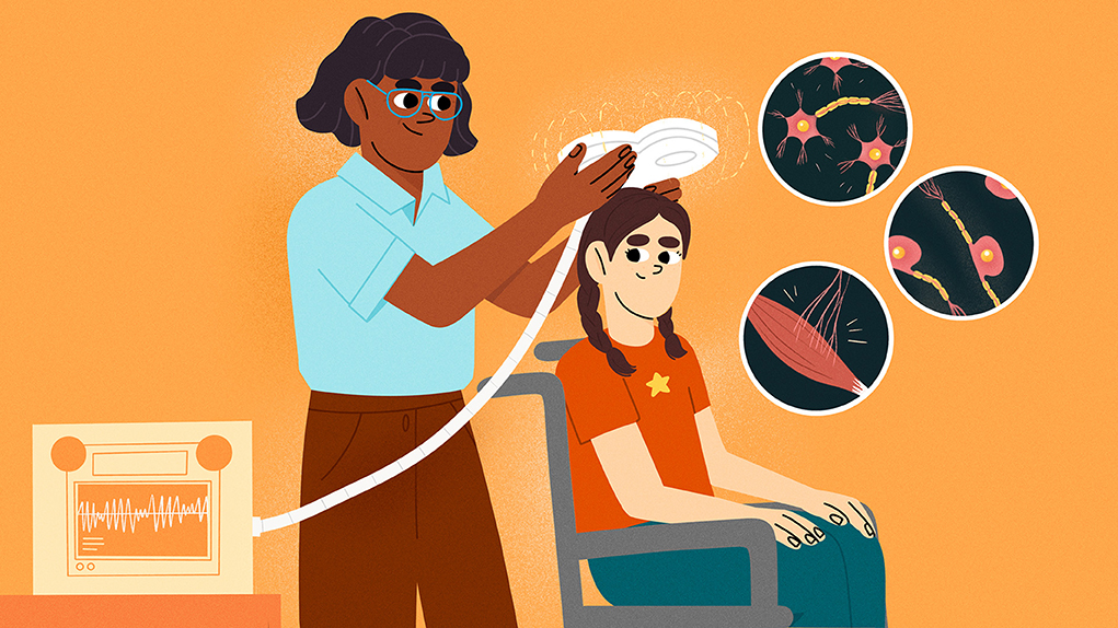
Whenever you read books, listen to music, or watch TV, your brain is using signals made from electricity and chemicals to help you understand the world around you. Your brain is full of cells called neurons that communicate with each other through these signals. Chemicals released from neurons transmit messages to their surrounding neighbors, telling the neighbors whether they should send a signal too. But it is electricity traveling down the length of the neuron that causes the release of those chemicals in the first place. Because electricity is involved in communication between neurons, scientists can use magnets to change the flow of electricity in the brain and explore how that affects behavior. A method called transcranial magnetic stimulation allows scientists who study the brain to stimulate the brain from outside a person’s head (through the skull). This gives scientists clues about brain functioning without requiring dangerous brain surgery!
Your brain is special; it is responsible for every aspect of your personality, it holds every memory you have ever created, it tells your body how to keep you alive, and so much more. Given its importance, it is not surprising, therefore, that it is well-protected, encased inside your skull. This protection, however, sometimes creates a challenge for cognitive and neuroscientists because it is hard to study something directly that you cannot touch or even really get close to. One common solution to this is to study the brain indirectly, by modifying its activity. That is, scientists can use special tools to disrupt the activity of neurons and then observe how this disruption leads to changes in a person’s behavior or their mental state. Neurons are specialized brain cells that carry information to one another using electricity and chemical messages (see Figure 1). Information is sent from one neuron to another using chemicals, but it is a small pulse of electricity that causes these messages to be sent in the first place. Because electricity is so crucial to this communication process, scientists can use magnets to disrupt the electricity in the brain and therefore alter the activity of the neurons. So how do tools like this work?
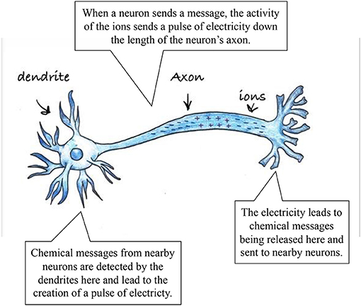
- Figure 1 - Neurons are cells that communicate using both electrical and chemical messages.
- Electrical messages travel down the length of the neuron and, when they reach the end, chemical messengers are released. The chemical messengers communicate with other nearby neurons in the network.
What is TMS?
Transcranial magnetic stimulation (TMS) is a method that safely delivers magnetic pulses through the skull to stimulate the neurons on or near the surface of the brain, changing their activity. When scientists change the activity of the brain using TMS, they also closely observe the participant’s behavior, or ask them how they are feeling, or both. When modification of brain activity leads to changes in behavior or mental states, that is a clue to scientists that the brain area they stimulated is important for (or involved in) that particular set of behaviors or feelings. This allows them to study the brain without being able to directly touch it. TMS is a safe and effective way to stimulate the brain that does not require surgery and has low risks for the person whose brain is being stimulated.
Long before we had tools like TMS, scientists could only study brains by dissecting them after a person died. But studying the brain after death does not tell scientists much about the activity of the brain when a person is alive. As surgery started to become safer, brain surgeons could open the skulls of their patients to examine the brains of living people. These studies gave doctors and scientists lots of information about what the brain does, but it is not the best method (obviously!) because it requires someone to undergo a major and potentially dangerous surgery. So, whenever possible, it is always best to study the brain without having to cut into anyone’s head!
By using TMS to study brain activity, scientists can learn which areas of the brain are responsible for various aspects of behavior, such as which areas are responsible for vision (for instance, seeing the shape of a candy bar), object recognition (realizing that the shape you see is a piece of candy), and motor control (using your arm and hand to pick up the candy and put it in your mouth!). Since TMS research began, it has been used as a way to treat depression, and it has also been used by laboratory scientists to understand brain functioning. TMS research can be incredibly beneficial for cognitive scientists; people who study the brain and behavior. For instance, by stimulating the areas of the brain that are responsible for language, cognitive scientists can better understand which parts of the brain are needed to understand language, and which parts are needed to produce language—it turns out that separate brain areas are responsible for these functions! Cognitive scientists can also do things like stimulate the premotor cortex—an area of the brain that helps people move. By doing this, they can begin to understand how the brain plans a movement and then performs that movement. But before we dive into what else scientists can learn by using TMS, we will tell you a bit more about how the brain works and how TMS affects it.
Finding Clues in the Body’s “Computer”
The brain is a complex organ that can be thought of as a bit like a computer. Just like a computer, the brain has multiple parts that work together to allow it to function properly. Inside the brain, there are billions of tiny neurons connected in many large networks. These networks allow one region of the brain to communicate with other regions of the brain, just like a computer might have wires allowing one part to “talk” to another. For a computer to function properly, all of its parts must be working and communicating. Your brain is the same! It needs its neurons and networks to communicate with one another to allow you to do all the amazing things you can do. When you interfere with one region of the brain using TMS, or interfere with the network sending signals from one region to another, the brain does not work in quite the same way. Sometimes that means that a person behaves differently than they would if their brain activity was not being disrupted. These changes in behavior allow cognitive scientists to act like detectives, using clues from behavior changes to figure out what parts of the brain are responsible for various aspects of behavior.
How Does TMS Work?
You may have heard that you should not place a strong magnet near your TV, your computer, or your phone. The reason for this is that the magnet can interfere with the flow of electricity through the device, sometimes disrupting its function temporarily or even damaging the device permanently. However, TMS is a safe and effective way to interfere with the flow of electricity in the brain, in a way that does not cause any lasting damage. Scientists can use TMS to selectively target one small region of the brain, but not in a way that would cause permanent damage or harm to someone.
To understand the logic of using TMS to study the brain, we can think of the brain as a computer once again. Imagine that you want to understand what each part of your computer does. One way to do that is to interfere with (unplug, disconnect, or break) certain parts of the computer to see what happens. If you unplug the fan, everything would get hot very quickly; if you disconnect the monitor, you would no longer see any picture; and so on. By interfering with selective parts of the computer, you can figure out what those parts do—a fan keeps the computer cool, a monitor shows you whatever you are working on. Well, TMS operates by the same logic, but we cannot easily and safely open up a person’s head to study the brain! Brain surgery is dangerous, and scientists are responsible for keeping their research participants safe and healthy. TMS can temporarily disrupt parts of the brain so scientists can figure out what those parts do, and this does not require surgery or put the participant at any risk.
In a typical TMS study, scientists place the TMS coil (which is a magnet shaped like a figure-8 or infinity sign) directly above the participant’s skull in the area the scientist wants to stimulate. For instance, the coil might be placed at the top of the skull if the scientist wants to stimulate the premotor cortex. After the coil has been placed in the right location, it is activated, which creates a small magnetic field that passes through the skull and into the outermost areas of brain tissue (Figure 2). This magnetic field creates an electrical current that affects neurons in the region of the brain near where the TMS coil was placed.
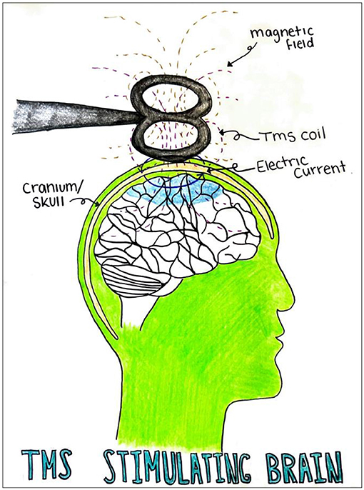
- Figure 2 - TMS stimulates the brain using a strong magnet.
- A TMS coil is placed directly above the brain area of interest, in this case the primary motor cortex. The area shaded in blue shows the part of the brain that is affected by the magnetic field, which disrupts the brain’s normal electrical currents. Because neurons are connected in networks, stimulating one area of the brain can also affect other areas.
To better understand how and why TMS is used by cognitive scientists, it is useful to learn about a specific study that used this tool. In 2002, some scientists wanted to understand the connection between two areas of the brain: the premotor cortex and the primary motor cortex [1]. These two brain areas are located next to each other, and areas of the brain that are close to one another usually (but not always!) talk to each other. Specifically, the premotor cortex seems to be responsible for planning movements, but the primary motor cortex tells the particular body parts to carry out the intended movement.
These scientists wanted to better understand how the connection between these two brain areas worked, and whether TMS stimulation to the premotor cortex would have an effect on the activity of the primary motor cortex. The research team found that TMS applied multiple times to the premotor cortex resulted in an effect on the neuronal activity in the primary motor cortex. This told the research team that stimulating the brain in one region can affect areas of the brain that are connected (through networks of neurons) to the part that was stimulated. Stimulating the premotor cortex changes what the primary motor cortex does, even though they are separate brain regions entirely!
How Can TMS Help People?
Scientists are using TMS to understand how the brain works, but this tool can also be used outside of research, to help people with various brain conditions. Two popular uses of TMS are therapy and rehabilitation. In therapy, TMS has been successfully used to treat people suffering from depression by stimulating the prefrontal cortex, which help us to control our emotions (among many other functions) [2]. TMS can be combined with medications, but it is often used as an alternative option for people who have not responded well to medications or who cannot take them.
Another important use of TMS is in rehabilitation. Various disorders (or injuries) of the brain or spinal cord can affect a person’s ability to move normally. TMS is now being used on the motor (movement) regions of the brain to help researchers learn more about repairing lost motor function. It is not uncommon for TMS to be used in combination with other treatments or other brain-stimulation methods [3].
We think that helping people is the future of TMS. Often, people think of scientists and medical professionals as two separate groups of people with very different goals: scientists try to discover how the brain works, while doctors try to help people by treating brain disorders. But the truth is that these two professions overlap a great deal and inform each other at all times. Without properly understanding how the brain works, doctors cannot hope to develop effective treatments for brain disorders or injuries. On the other hand, brain disorders and injuries often give scientists new questions to ask and new things to discover. TMS is an important tool not just because it is a safe and effective way to manipulate brain activity, but also because of what it can give us: a tool for discovery and a tool for treatment.
Glossary
Neurons: ↑ Specialized brain cells that send messages and work together to help us think, learn, and feel.
Transcranial Magnetic Stimulation: ↑ A procedure that uses magnetic fields (applied outside the skull) to stimulate neuronal activity within the brain.
Cognitive Scientists: ↑ People who study the brain, mind, and behavior, and explore mental processes such as thought, learning, memory, attention, and more.
Premotor Cortex: ↑ A region of the brain that helps plan and control movement.
Networks: ↑ Groups of neurons that work together and communicate regularly.
Therapy: ↑ A type of treatment designed to relieve a physical or mental disorder, disease, or injury.
Rehabilitation: ↑ The act of using training, medicine, or therapy to restore someone to a healthy state.
Prefrontal Cortex: ↑ A region in the front of the brain that helps us make decisions, solve problems, and plan ahead.
Conflict of Interest
The authors declare that the research was conducted in the absence of any commercial or financial relationships that could be construed as a potential conflict of interest.
References
[1] ↑ Münchau, A., Bloem, B. R., Irlbacher, K., Trimble, M. R., and Rothwell, J. C. 2002. Functional connectivity of human premotor and motor cortex explored with repetitive transcranial magnetic stimulation. J. Neurosci. 22:554–561. doi: 10.1523/JNEUROSCI.22-02-00554.2002
[2] ↑ Rizvi, S., and Khan, A. M. 2019. Use of transcranial magnetic stimulation for depression. Cureus 11:e4736. doi: 10.7759/cureus.4736
[3] ↑ O'Shea, J., and Walsh, V. 2007. Transcranial magnetic stimulation. Curr. Biol. 17:R196–R199. doi: 10.1016/j.cub.2007.01.030