
Abstract
Have you ever noticed that babies younger than 6 months only drink milk? This is because their digestive system is not quite developed enough to handle all the different foods and drinks that you can easily digest. You may have also noticed that babies cannot walk, talk, or read during that same time. This is partially because their brains also have not matured enough to let them do activities that you can easily do. The brain is growing very quickly at this time, so it is important to make sure babies are provided the best nutrition for brain growth. However, it is difficult to test babies; so our research looks at how different components in milk affect a piglet’s brain development, since pigs are much more similar to humans than you might have thought. This study shows that some components of milk may help the brain develop.
Nutrition Affects Brain Development in Babies
You may have heard that eating a diet full of fruits, vegetables, whole grains, dairy, and lean meats will help to make you strong, healthy, and smart. Children and adults who can eat solid foods have a large selection of items to choose from, but how do we make sure a baby, younger than 6 months, is getting a proper diet when he or she only drinks milk? By definition, mammals are a group of animals that have mammary glands capable of producing milk to nourish their babies early in life. While milk may look pretty simple to you, over 3,000 different components have been identified in human milk so far. Scientists have been working for over 100 years to better understand which components of milk are most important to ensure the best growth and development of babies. Although most babies drink milk, some babies are not able to drink it, and they rely on other sources of nutrition. This is another reason why it is important for scientists to figure out what components in milk help babies develop, so those components can be added to the diets of babies who cannot drink milk. While significant progress has been made, more work needs to be done to understand how nutrition affects the developing brain. This type of research will help ensure all babies have a chance at the best brain development, which will help them learn and remember things later in life.
You might wonder why nutrition is so important for a baby—they all drink milk, is that not enough? Scientists have discovered that some components of human milk vary over time and between mothers based on the mother’s genes and what she eats. Studying these changes over time and between different mothers has shown that certain nutrients like iron, choline, and DHA [docosahexaenoic acid (do·co·sa·hex·a·e·no·ic acid)] help the brain mature [1, 2]. While the impact of these dietary components has been extensively studied, there are many other components of milk that may have beneficial functions for brain development, but more research is needed to figure this out.
Therefore, our research focuses on studying components in milk, which may enhance brain development in babies. To do this, we first test dietary components in an animal, since it is often difficult to answer detailed scientific questions in human babies. We use piglets as a test animal because they have almost the same digestive system as human babies and a piglet’s brain grows similar to a human baby’s brain. Therefore, we can compare what might happen if a human were to drink these milk components by first giving these components to a piglet. In this study, we wanted to see how the addition of multiple components (called milk fat globule membrane, lactoferrin, and a prebiotic blend—you can find a description of each in Glossary) would affect the development of both the brain [3] and the digestive system [4]. In this article, we will focus on the techniques we used to understand how a diet containing these components influenced brain growth.
How Can the Piglet Help Us Understand How Milk Components Work?
In the field of nutrition research, it is important to study an animal that closely mimics what happens in our own bodies. By using an animal for these experiments allows scientists to look at many different examples of what happens, from body growth to how organs function, when different diets are eaten. Because piglets’ brains and digestive systems are similar to those of human babies, scientists can better understand the roles that dietary components play within the body and use this information to conduct similar studies in human babies. Animal research is important because a new dietary component must be proven to be safe and effective in animal experiments before it can be tested in humans.
When they were 2 days old, we placed 24 piglets in individual cages to make sure they received the diet we wanted them to eat. Of these pigs, 12 received the control (CONT) diet, which is a standard milk formula for piglets, much like human baby formula. The other 12 piglets received a diet containing the special ingredients (TEST), which was the CONT diet plus 3 dietary components mentioned above—milk fat globule membrane, lactoferrin, and a prebiotic blend. All piglets in the study received their diet five times a day from 2 to 31 days of age. Because the TEST diet only differed from the CONT diet by having the three additional components, we compared piglets fed these diets to see how the TEST diet may have changed how the piglets’ brains developed.
Fun Fact
The pig brain is 1/10th the size of the human brain, but it looks like the human brain and grows in the same way that the human brain grows!
Magnetic Resonance Imaging (MRI) in Piglets
When the piglets were 30 days old, they were subjected to MRI using the same type of scanner that is used for humans. A person can lay in an MRI machine to safely have images taken of their brain without their body being touched in any way. An MRI machine has a strong, but safe, magnetic field, and the atoms in the bodies align with this field when a person lays in the machine. The MRI machine sends signals that knock these atoms over, and as the atoms come back in line with the field, the MRI sees this and generates an image of the brain from the data. These MRI scans helped us to compare the brain growth of piglets on the TEST and CONT diets to see if they were different. By using the same type of MRI scanner for pigs and humans, we also got information that helps to inform scientists of what they might expect to see if a human baby were to receive a diet similar to the one the piglet received (Figure 1). You can see in Figure 1 that piglet and human brains have similar structure because they both have the bumps, also called gyri, and grooves, also called sulci.
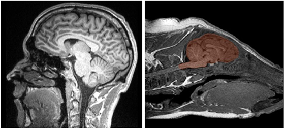
- Figure 1
- The image on the left is a magnetic resonance imaging (MRI) image of a human brain. On the right is an MRI image of a piglet, with the brain shaded in red. Pigs and humans both have gyri (bumps) and sulci (grooves) on the outer surface of their brains, which is one way that human and piglet brain development is very similar.
Fun Fact
If you have ever had an MRI or seen one on TV, you might have noticed it was noisy. This is because of the signals sent from the MRI machine that help to create the image of the brain.
Measuring Myelination in the Brain
The brain is made of cells called neurons. Each neuron has a cell body and a long, tail-like structure called an axon, which is used to transfer information from one neuron to another. Throughout the first few years of life, the brain undergoes a process called myelination. If you think of an axon in the brain as an electrical cord, the rubber coating on the electrical cord would be what scientists call myelin. As we get older, the myelin sheets get thicker around the axon to help it work more efficiently. When the protective covering of an old electrical cord starts to come off, the appliance does not work properly because electricity is not efficiently conducted through the wire. This would be similar to an axon that is not properly myelinated, making it work less efficiently. Most myelination occurs when we are babies and young children, so it is important for scientists to understand how early-life nutrition helps the process of myelination. We need properly myelinated axons to perform everyday functions, so making sure that the axons are myelinated early in life is very important.
One way to analyze the MRI data is through a process called diffusion tensor imaging (DTI). This MRI technique assesses how fast and where water molecules are moving in the brain. By knowing how the water moves, we are able to understand how neurons are aligned and how myelination is occurring in the brain; these are important ways of measuring brain development. In general, it is better to have neurons lined up in the brain so that they are all going in the same direction, like uncooked spaghetti noodles that all line up together, rather than neurons that are crossing all over each other, like a giant bowl of cooked spaghetti.
In our study, we used DTI to observe differences in something called radial diffusivity (dif·fu·siv·i·ty) between piglets on each of the diets and to help determine how much myelin had developed in their brains. Radial diffusivity is how fast water is moving perpendicular to (or across) the lined-up axons. When babies are born, they do not have much myelin surrounding their axons, so water can flow out of the axons fairly easily; this would result in a high radial diffusivity measure (Figure 2). However, as babies’ brains develop, myelin wraps around the axons and there is less room for water to move out of the axons, resulting in lower radial diffusivity. When compared with CONT piglets, the piglets that received the TEST diet had lower radial diffusivity measures in a brain region called the internal capsule. This indicates that the TEST piglets likely had more myelination in the internal capsule compared with piglets on the CONT diets, meaning the diet may have enhanced brain development in the internal capsule. This brain region is important because it is one of the first to develop in babies. When you think of a baby, some of the first things he or she can do are reach for objects and touch them—this is because their internal capsule helps with motor movement and with sensing the environment.
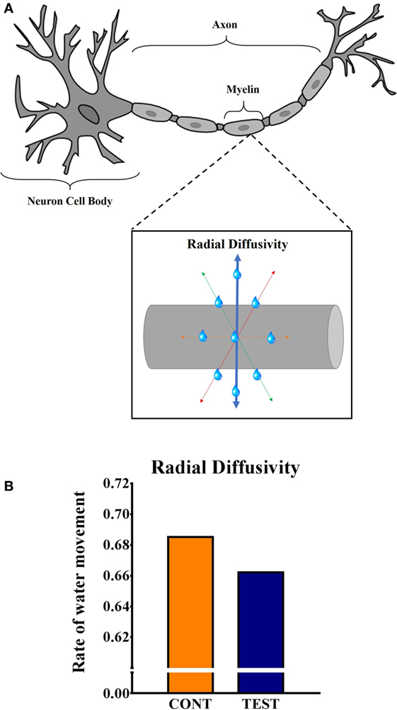
- Figure 2
- A. This is an illustration of a neuron, showing myelin wrapping around the long part of the neuron called the axon. When a neuron cell body receives information from other neurons, the cell body processes that information and sends a new message along the axon to reach other neurons within the brain or in other sites within the body. The box shows an enlarged image of part of the axon. As you can see in the box, water molecules (blue drops) can move in all directions. Radial diffusivity is water movement across the axon (along the blue line). B. Piglets that received the TEST diet had lower measures of radial diffusivity in a part of the brain called the internal capsule compared with CONT piglets. This likely means the TEST piglets had increased myelination in the internal capsule.
Fun Fact
You may have heard someone say the brain is mostly made of fat. This is true, and myelin is a big part of what makes the brain fatty. Now do you see why it is important for babies to have healthy fats in their diet?
Looking at Differences in Gray Matter Concentrations
Another way we analyze the MRI data is called voxel-based morphometry (VBM). All of the MRI images are made up of what are called “voxels”; you can think of this as the brain being digitally split into lots of tiny three-dimensional cubes that are 0.7 mm on each side. Each cube contains brain tissue that may be gray matter, white matter, or sometimes a combination of both (Figure 3). Gray matter is part of the brain that mainly contains neuron cell bodies, which are tightly packed together, making it appear gray in color. White matter areas in the brain are composed mainly of myelin, and it appears white because of the large amount of fat in the myelin. To analyze the differences in the brain using VBM, we first “segment” or separate each piglet’s brain into an image that contains only gray matter cubes and one that contains only white matter cubes. We can do this easily with a computer program. We are then able to compare the gray or white matter images from piglets on different diets to see if there are areas of their brains with different amounts of gray or white matter tissue.
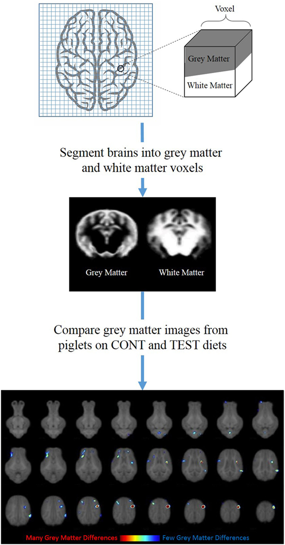
- Figure 3 - This is a flow chart of how the voxel-based morphometry data were generated.
- The piglet brain was digitally split into many three-dimensional cubes called voxels. These cubes have gray matter, white matter, and sometimes both tissue types within them. By using a computer program, we then separated the cubes into those that contained gray matter and those that contained white matter. From these images, we compared the amount of gray matter in the brains of piglets on the two different diets. At the bottom of this figure, you can see a normal piglet brain separated into many layers from the bottom of the brain (top left image) to the top (bottom right image). All the places where you see colored spots are areas where the CONT piglets had more gray matter than the TEST piglets. The red color indicates an area that has much more gray matter in the CONT than in the TEST pigs. The blue color indicates an area where there is only a small difference in the amount of gray matter between the two groups. This might mean that the brains of CONT piglets did not undergo pruning at the same time as the TEST piglets, indicating a change in the pattern of brain development.
By performing the VBM analysis on the piglets from this study, we observed differences in the distribution of gray matter between pigs fed the two diets. It was interesting that, compared with TEST piglets, piglets that received the CONT diet had more gray matter in the cortical regions, which is where many of the neuron cell bodies are located. You might think that more is better when it comes to the brain, but early in life the brain produces many more neurons than we actually need as adults. The brain then goes through a process called “pruning,” when the neurons that are not needed are removed to make room for neurons that we do need. Because the piglets on the TEST diet had less gray matter compared with the CONT piglets, we speculate that the TEST piglets underwent pruning of neurons at an earlier age, which could mean the TEST diet helped the piglets’ brains to develop faster. In addition, the brain region where we saw differences in gray matter is located near the areas that control movement and sensory information. Can you imagine why this would be helpful for human babies?
Fun Fact
Just after birth, our brains grow faster than they grow at any other time in our life. This is why it is very important to have good nutrition when we are babies, so that we do not slow our brain growth.
Why is This Work Important?
When we look at the brain imaging outcomes from this study, a very interesting story emerges. Both of the brain imaging techniques tell a similar story, indicating that TEST piglets had advanced brain growth compared with CONT piglets. Studies in human babies have also shown that the addition of milk fat globule membrane to a baby’s diet can help the brain develop [5, 6]. This might be because milk fat globule membrane contains a molecule called sphingomyelin (sfing·goh·mahy·uh·lin), which is required for the production of myelin. These human studies support our findings that feeding piglets a diet containing milk fat globule membrane helps their brains develop. It is important to remember that the piglets’ TEST diet also contained lactoferrin and a prebiotic blend, but more research needs to be done to understand what role these components may play in brain development. Now that we know a diet containing these compounds helps piglet brain growth, and more research needs to be done in humans to see if the same is true for human baby brains. Before reading this paper, you may have looked at a baby drinking milk and not thought any more about it. Now you know that not only is the milk important for the baby but also specific components in the milk help the baby to think, move, and develop properly!
Ethics Statement
The study was approved by University of Illinois at Urbana-Champaign Institutional Animal Care and Use Committee.
Glossary
Milk fat globule membrane: ↑ A component of milk that is made up of both proteins and specialized fats.
Lactoferrin: ↑ A protein in milk that gets rid of bad bacteria and therefore helps babies to not get sick.
Prebiotic blend: ↑ A combination of carbohydrates that help good bacteria grow in the digestive system.
Magnetic resonance imaging: ↑ A pain-free test that uses a combination of radio waves and a strong magnetic field to produce pictures of tissues within the body.
Myelination: ↑ The process of wrapping myelin around an axon to help it function better.
Myelin: ↑ A combination of fats and proteins that form in a sheet around the axon to help make sure the neuron is able to communicate properly with other neurons.
Diffusivity: ↑ A measure of how fast water molecules move around.
Internal Capsule: ↑ Part of the brain that connects areas responsible for generating movement and sensing the environment with the rest of the body, and this region of the brain is one of the first to mature in babies.
Gray matter: ↑ Part of the brain that mainly contains neuron cell bodies. This area appears gray because there are many cells bunched together making it appear dark.
White matter: ↑ Areas of the brain that contain myelin that wraps around the axons of neurons. This area appears white because it is made of many fat molecules.
Sphingomyelin: ↑ A special type of fat that helps to form the myelin that wraps around axons.
Conflict of Interest Statement
BB and RW are employees of Mead Johnson Nutrition. SD and RD have received grant funding from Mead Johnson Nutrition. Authors declare no other potential conflict of interest.
Original Source Article
↑ Mudd, A. T., Alexander, L. S., Berding, K., Waworuntu, R. V., Berg, B. M., Donovan, S. M., et al. 2016. Dietary prebiotics, milk fat globule membrane, and lactoferrin affects structural neurodevelopment in the young piglet. Front. Pediatr. 4:4. doi:10.3389/fped.2016.00004
References
[1] ↑ Georgieff, M. K. 2007. Nutrition and the developing brain: nutrient priorities and measurement. Am. J. Clin. Nutr. 85:614–20.
[2] ↑ Innis, S. M. 2008. Dietary omega 3 fatty acids and the developing brain. Brain Res. 1237:35–43. doi:10.1016/j.brainres.2008.08.078
[3] ↑ Mudd, A., Alexander, L., Berding, K., Waworuntu, R., Berg, B., Donovan, S., et al. 2016. Dietary prebiotics, milk fat globule membrane and lactoferrin affects structural neurodevelopment in the young piglet. Front. Pediatr. 4:4. doi:10.3389/fped.2016.00004
[4] ↑ Berding, K., Wang, M., Monaco, M., Alexander, L., Mudd, A., Chichlowski, M., et al. 2016. Prebiotics and bioactive milk fractions affect gut development, microbiota and neurotransmitter expression in piglets. J. Pediatr. Gastroenterol. Nutr. 63(6):688–97. doi:10.1097/MPG.0000000000001200
[5] ↑ Tanaka, K., Hosozawa, M., Kudo, N., Yoshikawa, N., Hisata, K., Shoji, H., et al. 2013. The pilot study: sphingomyelin-fortified milk has a positive association with the neurobehavioural development of very low birth weight infants during infancy, randomized control trial. Brain Dev. 35(1):45–62. doi:10.1016/j.braindev.2012.03.004
[6] ↑ Timby, N., Domellöf, E., Hernell, O., Lönnerdal, B., and Domellöf, M. 2014. Neurodevelopment, nutrition, and growth until 12 mo of age in infants fed a low-energy, low-protein formula supplemented with bovine milk fat globule membranes: a randomized controlled trial. Am. J. Clin. Nutr. 99(4):860–8. doi:10.3945/ajcn.113.064295