Abstract
Imagine an expert piano player’s hands as they fly across the piano keys. Have you ever wondered how it is possible to control such complicated and precise movements? The brain controls our movements and processes all the information we receive from the entire body as we interact with the world. We perform many complex movement-related tasks, and our brains manage them all the time without us even noticing! To manage the whole body efficiently, the brain is organized so that specific brain areas control or gather information from specific body parts. The parts of the body that perform the most complex tasks are assigned more space in the brain. Can you think of a body part that is responsible for very complex movements? What would happen in your brain if you could no longer move that body part?
The Brain Is Organized for Maximum Efficiency
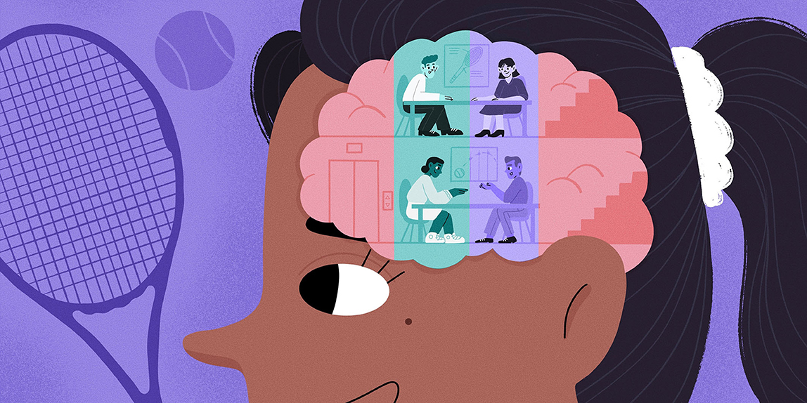
Our brains interpret the world around us, think, imagine, control our movements, and much more (see this Frontiers for Young Minds article). This seems like a lot of work for one small organ! How can the brain control so many different tasks? The best way to deal with many duties is to distribute the work, and this is how the brain does it (read more here). Specific parts of the brain are responsible for processing what we see, controlling how we speak, regulating how we move, and so on. In a busy office building, people who need to speak to each other regularly will often have their desks near each other, but people who have very different jobs and tasks can work on separate floors. It would not make sense for members of the same team to go up and down the stairs all day when they could just sit together! Putting people with similar tasks near each other allows them to work faster. The brain uses a similar strategy. Similar brain functions are controlled by brain regions that are close to each other. This means that the brain regions that often work together can communicate quickly and efficiently.
The Brain’s Movement Control Center
Several brain areas are involved in movement, and the motor cortex is a very important one (Figure 1). Cortex means “bark,” like the bark of a tree. Just like tree bark, the cortex is the outer layer of the brain. “Motor” means movement, so the motor cortex is the part of the brain that tells the muscles to move the body. Think of all the movements you make when you play an instrument or a sport—the brain must control many muscles all at once (read more about it here)! This is quite a complex task for the motor cortex. How do you think it handles this problem? Just like the entire brain is organized by function, the motor cortex is organized so that certain locations control certain body parts (Figure 1). One half of the body is represented in one half (hemisphere) of the brain, while the other half is represented in the other hemisphere. Interestingly, the right hemisphere controls the left side of the body and the left hemisphere controls the right.
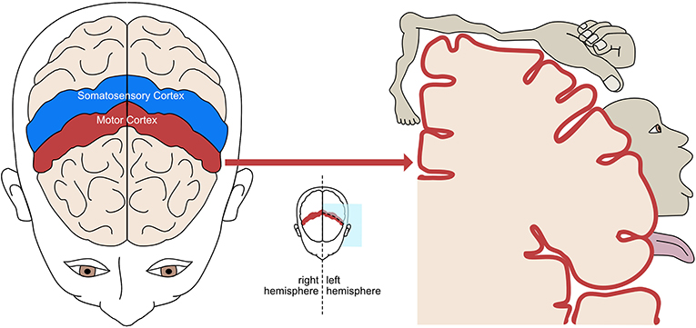
- Figure 1 - The motor cortex (red) and somatosensory cortex (blue) are located in the central part of the brain, stretching from ear to ear.
- Both are organized so that each part of the body maps to a specific part of each cortex. In the cross-section on the right, the cartoon person shows the approximate location of each body part and the brain space it takes. Body parts with more complex functions, like the hands and mouth, have a bigger representation in the brain than parts with simpler functions, like the feet.
The somatosensory cortex (Figure 1) is also very important for movement. “Soma” means body, and “sensory” refers to sensations that help us understand and interact with the world around us, like touch, temperature, and pain. For example, we can feel cold snow on our hands when we touch it, or feel pain if we fall and scrape our knees. And if we close our eyes and move our bodies, we still know where our arms and legs are, even if we can not see them. These sensations come from sensory receptors, located in the skin and muscles, that measure what we feel and send this information to the brain. So, the somatosensory cortex is the part of the brain that processes and interprets sensations coming from the body. The motor and somatosensory cortices are strongly connected and communicate constantly. Their communication must be very efficient because every movement causes somatosensory sensations, and these sensations affect how we move next. The organization of the somatosensory cortex is similar to that of the motor cortex—specific locations process the sensations coming from certain body parts.
The Homunculus: A Map of the Body in the Brain
To control the body, the brain must tell specific body parts to move. To help with this, there is a “map” of the body in the motor cortex. Body parts that are attached to each other, like the hand and arm, are positioned near each other in this brain map. But this efficient organization does not end here. Do you use all your body parts equally, or are some parts capable of more precise actions? Think about your hands and feet, for example. Could you thread a needle with your toes? Some body parts have more difficult jobs, so they require more brain resources. The brain assigns bigger areas for tasks that are more demanding. As a result, the area of the brain corresponding to the hands is bigger than the area corresponding to the feet. The body map in the brain does not match the physical size of each body part, but instead matches how important and precise each body part’s job is. If we drew each body part with a size proportional to the space it is assigned in the brain, the resulting picture would be a strange-looking body with very large hands and very small legs. This figure is called a homunculus [1], which literally means “little person” (Figure 2).
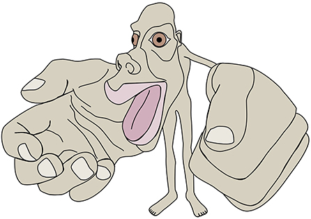
- Figure 2 - The motor homunculus is the name for a “little person” represented by the brain areas assigned to each body part in the motor cortex.
- Body parts, like the hands and mouth, that are required to make very complex and accurate movements are given more brain area. That is why the homunculus looks different from an actual human body, with very big hands and mouth and small feet.
The somatosensory cortex also has a homunculus organization. The parts of the cortex that control the movement of a specific body part and feel sensations from that part are close together, so they can communicate quickly. For example, the hand area of the motor cortex is right next to the hand area of the somatosensory cortex. Like the motor homunculus, the sensory homunculus also has very big hands. This is because we have many sensory receptors in our hands since we explore the world more with our hands than with our feet. All mammals have a similar brain organization that matches the use of their body parts. For example, mice explore the world with their whiskers, so they have a large brain area dedicated to them [2].
Why Is the Homunculus Organization Useful?
The collaboration between senses and movement is very important. Try closing your eyes and lifting a bottle full of water. Then do the same, but with an empty bottle. The muscle force needed will be different, but you can quickly adjust to the right amount of force just by feeling the bottle. This is because the motor and sensory homunculi are a great team and talk to each other constantly. Your motor cortex sends instructions to your muscles to wrap your fingers around the bottle. When your hand touches the bottle, sensory receptors in your hand record information about the sensations and send it to the somatosensory cortex. The hand area in your somatosensory cortex processes the information and sends it to the hand area in the motor cortex. This information helps you decide how much to squeeze or release. This happens so quickly you do not even realize it, which is really important for fast and accurate control of your movements. In some people with motor disabilities, the communication between the sensory and motor cortices does not work very well, which makes every movement more difficult.
We mentioned that the homunculus has very big hands. Can you imagine another body part that will be very big in the homunculus? Think about the importance of your mouth for speaking and eating. Even just by imagining your mouth, the mouth area in your brain is activated—you can activate your motor cortex just by thinking about moving! For example, if you play with a ball and touch it with your hands, the hand areas in your brain will activate. Later, if you think about touching the ball, the same brain areas will activate even if you are not touching the ball (Figure 3). When you just imagine the movement, other parts of the brain get involved to prevent your muscles from moving.
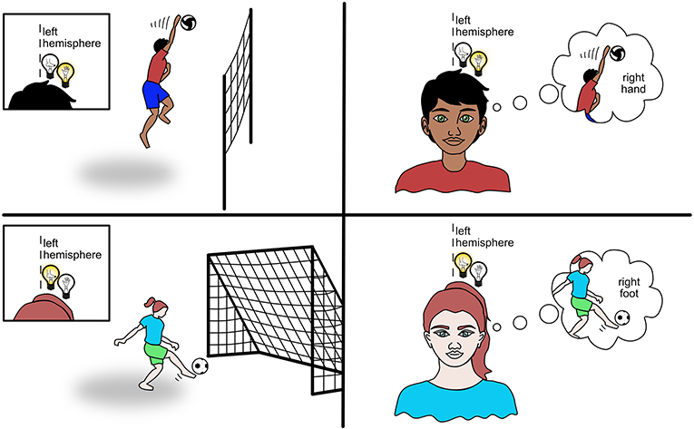
- Figure 3 - When you use your hands or feet, different areas of the brain will activate.
- If you touch a ball with your hand like the volleyball player, the hand area in the opposite brain hemisphere activates. If you touch a ball with your right foot like the soccer player, the foot area in the left brain hemisphere activates. The same area of your brain that activates while hitting the ball with your hand or foot also activates when you think about hitting the ball with your hand or foot, as you can see on the right side.
The brain activation that happens when we imagine movements is very powerful because it helps us learn and it can even speed up recovery from motor impairments, like those caused by a stroke (read more about it in this Frontiers for Young Minds article). A stroke occurs when a part of the brain gets damaged because a blood vessel supplying oxygen and nutrients is blocked or damaged. If a stroke occurs in part of the brain which controls movement, this may result in inability to move some body parts. If this damaged brain area is on the left hemisphere, the right side of the body will be impaired and vice versa. If the hand and/or the leg area is affected the corresponding body part will be affected. Knowledge about the homunculus can help doctors design therapies to train specific parts of the body after brain injury or stroke (see this article) and can help neurosurgeons plan brain surgeries.
The homunculus not only helps us to control our own bodies, it also helps us to understand the movements of other people. When you see someone moving, you can imagine what it feels like, because we all have a similar homunculus. When you see someone moving their hand, the hand area in your brain will also activate. This helps us to copy the movements of others and to learn by watching. It also makes us feel more socially connected because if you see someone scrape their knee, you can imagine how painful it is and come to give them a hug.
Can the Homunculus Change?
The homunculus is so important and similar across people that it is already present when we are born [3]. But this does not mean it cannot change. The homunculus can adapt when we need to use our bodies differently. We can see an example of this in a person who loses their whole hand in an accident. Scientists found that the motor area that normally activates during hand movements becomes active during arm movements instead [4]. So, it looks like the homunculus’s arm gets bigger and its hand disappears, since there is no hand to control. But this process can also be reversed! Sometimes, a person who has lost a hand can have a replacement surgically attached. Thanks to this very special operation and a lot of training, the person can learn to control the new hand fairly well. The homunculus’s hand area, which was taken over by the arm area, can again become active during hand movements. So, our brains have an amazing ability to adapt to meet the changing needs of our bodies. This is known as brain plasticity. We still have a lot to learn about how brain plasticity works, but it is good to know that our brains are capable of such impressive changes with training and practice. You have probably heard many times that “practice makes perfect”. That saying refers to how we learn and improve our skills due to brain plasticity. So, if you want to learn a new skill or recover from an injury, the best thing to do is train and practice!
In summary, the functions of the brain are organized in space, so they work together at their best. Functions related to body movements and sensations are localized in specific areas of the brain depending on the specific area of the body involved. The homunculus is a way to describe how the body is represented in the brain. This representation may change due to brain plasticity, which is critical for learning new motor skills and recovering from injuries.
Acknowledgments
This project has received funding from the European Union’s Horizon 2020 research and innovation program under the Marie Sklodowska-Curie grant agreement No 846679 (INFANTPATTERNS) and the Life Science Engineering Area of Advance at Chalmers University of Technology.
The authors would like to thank Tomoki Arichi and James Arichi for reading and commenting on the article.
Figures 1, 2 in this article contain content which is adapted from the following images made publicly available through Creative Commons:
• https://en.wikipedia.org/wiki/Cortical_homunculus#/media/File:Motor_homunculus.svg
• https://commons.wikimedia.org/wiki/File:Sensory_and_motor_homunculi.jpg.
Glossary
Motor Cortex: ↑ Brain area involved in planning, controlling, and performing movements.
Hemisphere: ↑ Literally, half of a sphere. The brain is divided into two almost symmetric halves, called brain hemispheres.
Somatosensory Cortex: ↑ Brain area responsible for sensations of touch, pain, temperature, and body position, which come from our skin and muscles. “Soma” means body, so these are body-related sensations.
Sensory Receptors: ↑ Structures that receive somatosensory information from the skin and muscles and send it to the brain.
Homunculus: ↑ Means “little person” and refers to the representation of the whole body in the brain, based on the complexity of movement or sensation.
Stroke: ↑ Reduction of the blood supply to a part of the brain causing damage.
Brain Plasticity: ↑ The brain’s ability to change and adapt as a result of experience.
Conflict of Interest
The authors declare that the research was conducted in the absence of any commercial or financial relationships that could be construed as a potential conflict of interest.
References
[1] ↑ Penfield, W., and Boldrey, E. 1937. Somatic motor and sensory representation in the cerebral cortex of man as studied by electrical stimulation. Brain. 60:389–443. doi: 10.1093/brain/60.4.389
[2] ↑ Zimmer, C. 2013. Mouseunculus: How the Brain Draws a Little You. National Geographic Available online at: https://www.nationalgeographic.com/science/article/mouseunculus-how-the-brain-draws-a-little-you (accessed November 08, 2022).
[3] ↑ Dall’Orso, S., Steinweg, J., Allievi, A. G., Edwards, A. D., Burdet, E., and Arichi, T. 2018. Somatotopic mapping of the developing sensorimotor cortex in the preterm human brain. Cereb. Cortex 28: 2507–15. doi: 10.1093/cercor/bhy050
[4] ↑ Giraux, P., Sirigu, A., Schneider F., and Dubernard, J.-M. 2001. Cortical reorganization in motor cortex after graft of both hands. Nat. Neurosci. 4:691–2. doi: 10.1038/89472