
Abstract
We humans have more than 600 muscles in our bodies! You constantly use your muscles to eat, breathe, make faces, and run around! How many muscles do you think a person uses to play the piano, dance the macarena, or play basketball? How do you think the muscles get the message to move? We cannot see our muscles under the skin, so it is easy to forget about them. If you make the “strong arm” pose by flexing your elbow and clenching your fist, you might see a muscle bulge. Or if you get a lot of exercise, your muscles might get tired or sore. Would you like to be able to see what your muscles are doing under your skin? If you could listen really closely, do you think muscles make any sound? In fact, with some special recording equipment, it is possible to see and hear what muscles are doing!
Intro To Human Muscles
When you intentionally move your body, you use your skeletal muscles. Skeletal muscles got their name because most of them attach to the bones of the skeleton. The skeleton provides the structure of the body and the muscles move the skeleton around. Other kinds of muscle make movements you do not think about or control. For example, the heart is a muscle that works non-stop, moving blood throughout the body. You also have smooth muscles such as those that change the size of the iris (the colored part of the eye) or give you goose bumps when it is cold. In this article, we will focus on the skeletal muscles.
Skeletal muscles are made up of many individual muscle cells. These cells are called muscle fibers because they are shaped like long, thin strings. Muscle fibers are bundled together in various ways to form many different muscle shapes. Some muscles are shaped like circles, like the mouth, and some are shaped like ribbons, fans, or feathers (Figure 1)! Each muscle’s shape and the way it attaches to the skeleton give it a special role in moving the body. Pairs of muscles are responsible for moving a joint (like the elbow) in opposite directions. For example, the biceps moves the forearm toward the upper arm, while the triceps moves it the opposite way. This is because muscles can only pull and not push! Muscles pulling in opposite directions on the same joint are called antagonists.
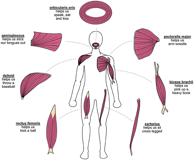
- Figure 1 - Muscles have many different shapes and attach to the body in many different ways, which allows each muscle to do its unique job.
- The yellow structures are called tendons, which attach skeletal muscles to the skeleton. Try putting your hand over each muscle in the picture while you perform the indicated action. Can you feel your muscles moving? The genioglossus is covered by another muscle, but you might still feel its movement by placing your hand under your chin. All these actions require many muscles working together, so you can probably feel other muscles moving too!
Muscle Fun Facts
Can you guess which muscles in your body are:
The strongest?
The most active?
The longest?
The smallest?
The weirdest?
Eating is critical for staying alive. This is why the strongest muscle in the body is the chewing muscle! It is called the masseter, which means “to chew” in Greek.
Many muscles in the body are active almost all the time. The eye muscles are an example. If you pay attention to your eyes, you will notice they are constantly making tiny movements. This happens even while you are asleep and dreaming!
The longest muscle in the human body is the sartorius (Figure 1). This is the muscle that is activated when you sit cross-legged. In fact, its name means “tailor’s muscle,” in reference to the cross-legged position in which tailors used to sit.
The smallest muscle is the stapedius, which stabilizes the stapes in the ear, the smallest bone in the human body. Despite its small size (about the thickness of a coin), this muscle is very important because it helps to protect the ears from loud sounds.
The tongue is a set of muscles with quite a weird organization! Most skeletal muscles attach to bone or tissue at both ends, but the tongue has one end completely free. This allows you to move your tongue in all directions, to help you speak, chew, taste, and swallow.
Muscle Movement
The brain tells the muscles to move, then the muscles move the skeleton. But how does this actually work? The answer is electricity, just like the electricity that makes the lights work! The brain sends electrical messages to the muscles through a system of biological wires called neurons [1]. A neuron that carries messages about movement is called a motor neuron. The electrical messages are called signals because they contain important instructions for the body. These signals are tiny compared to the electricity in a lightbulb, so you do not feel any shock. Motor neurons leave the spinal cord and split into many branches. Each branch activates one muscle fiber. So, each motor neuron activates a group of muscle fibers all at once. One motor neuron and its group of muscle fibers is called a motor unit. When the signal reaches the muscle fibers, the muscles activate and pull on the skeleton. When enough fibers are active, the skeleton moves (Figure 2)!
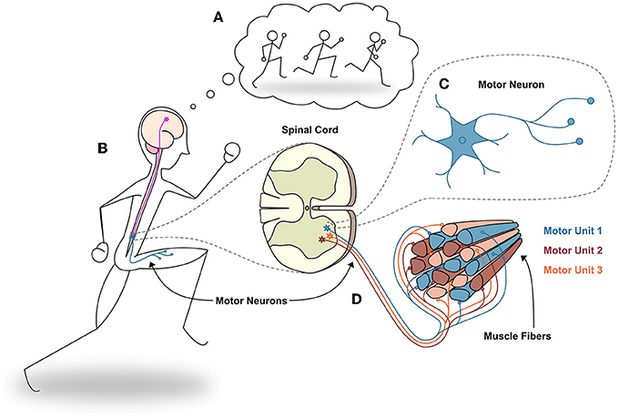
- Figure 2 - Activating the leg muscles.
- (A) We plan a movement. (B) Upper motor neurons send the signal from the brain to the spinal cord, and lower motor neurons send the signal from the spinal cord to the muscle. (C) A close-up of a motor neuron. (D) Each lower motor neuron activates a group of muscle fibers, making them pull. Each lower motor neuron and its group of muscle fibers is called a motor unit. Motor units are like miniature muscles that make up a whole muscle. If enough motor units are activated, your muscles move your body!
The brain uses three tools to control the strength, speed, and coordination of movements. First, motor units are only activated when they are needed. Stronger movements need more motor units, and weaker movements need fewer. Second, the brain controls the firing speed of motor units. When a motor unit activates, it sends one very fast electrical signal, so we say it “fires.” To make stronger and faster movements, the motor unit fires more quickly. Last, to control the direction and complexity of movements, the brain can activate many muscles in combination. Imagine the millions of signals that are firing all over your body when you dance or run! If you could hear and see them, you would be crackling like lightning!
How Can We Hear and See Muscles?
Electromyography (EMG) is the method scientists use to record the electrical activity of muscles (Figure 3). First, EMG detects the muscle signals. To detect the signals, doctors, or researchers place two electrodes on the skin over the muscle, to measure how the electrical signal flows from one point to another. Electrodes are metal plates that move electricity from the body to the recording equipment, so it can be measured. These signals are very tiny, so they must be amplified so we can see and hear them. If you have heard an electric guitar, you have probably heard the effects of an amplifier. Without an amplifier, an electric guitar makes a tiny sound. The amplifier increases the strength of the sound so you can hear it during concerts. Similarly, amplifiers strengthen the muscle signals so they can be seen on a computer. Then we can view and study the signals. Electrodes on the skin cannot tell motor units apart—we see the signals added together as in Figure 3A.
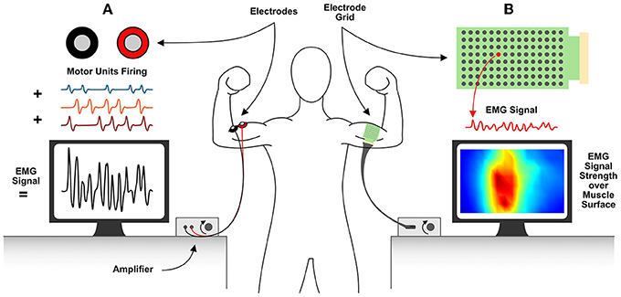
- Figure 3 - EMG is the system used to record the electrical activity of muscles.
- The signal is detected with the electrodes, strengthened by the amplifier, and studied on the computer. (A) The colored signals show the activity of each motor unit. The electrodes detect all motor unit signals as if they are added together, which you can see in black on the computer screen. (B) Many electrodes record EMG signals in a grid pattern and are processed on the computer to map the EMG activity over the muscle surface.
Muscle Sounds
With amplifiers and special electrodes, researchers can hear muscles! If you have heard popcorn popping, you know what muscles sound like! Imagine that every pop is the sound of a motor unit firing. With popcorn, at first it pops very slowly and you can hear individual pops with time in between pops. This is what your muscles sound like when you start using them. Then popcorn pops like crazy, and you cannot tell one pop from another! This is what your muscles sound like when you make a strong movement. Click Video 1 to listen to the sound of motor units firing: one slowly, one quickly, and then both together [2]. Can you tell them apart?
- Video 1 - When we use an amplifier, we can hear individual motor units firing. Each time a motor unit fires, it sounds like a pop. Here, you can hear the amplified sound of two motor units firing. First, you hear one motor unit firing slowly, then another one firing quickly, then both firing at the same time. Imagine what it sounds like when there are 10 or 100s of motor units firing at the same time!.
Muscle Maps and Movies
To see what is happening in a large area of muscle, we can make a map! Our eyes cannot see electricity, so we use some technology tricks. We arrange many electrodes in a grid pattern and place them on the skin over a muscle (Figure 3B). If one area of the muscle is more active than another, the electrodes above it detect a stronger signal. We assign the signal strength at each electrode to a rainbow of colors, between blue and red. Blue is for weak signals, red is for strong signals, and green and yellow are in between.
As the person wearing the electrodes moves, the muscle map changes. To see how muscle activity changes over time, we have made a muscle movie [3]! Click Video 2 to see the muscle activity in the biceps while a person is flexing the elbow, as shown in Figure 3 [4].1 If you make this pose and activate your biceps, you will notice that the skin barely moves, but the video shows that the muscles are very active!
- Video 2 - This video shows a muscle movie created from EMG recordings taken while a person flexed their elbow, like in Figure 3. A grid of 120 electrodes (8 rows and 15 columns, spaced 1 cm apart) was placed over a person’s biceps brachii muscle (see Figure 1) during the movement. Signal strength was estimated for each EMG signal and shown in a range of colors, where weak signals are blue and strong signals are red, like in Figure 3B. A muscle movie makes it very easy to see which parts of a muscle are most active during a movement.
What Can We Do With Muscle Signals?
Scientists and doctors can do a lot of useful things with EMG recordings! For example, to treat problems with muscles or nerves, we need to know what is wrong and where. For over 70 years, doctors have used EMG to diagnose nerve and muscle malfunctions. They do this by comparing EMG signals from healthy and sick patients. Also, scientists, doctors, and engineers use EMG to create muscle-controlled prostheses [5]. A prosthesis is a device that replaces a missing function in a person’s body. If someone loses a hand, he or she might receive a hand prosthesis to replace it. Using EMG signals recorded from the arm, a person with a muscle-controlled hand prosthesis can move it in simple ways, by activating the arm muscles.
EMG can teach us a lot about how the brain controls the body. We have many useful methods for recording and studying EMG data. Learning to use these methods and inventing new ones will help us find creative new ways to help people who have nerve, muscle, or movement problems. This is the job of doctors, physiotherapists, biomedical engineers, and movement scientists.
The next time you eat, run, dance, or play an instrument, remember the pop and color of your electrified muscles! Can you think of more ways to use EMG to help people?
Funding
This project has received funding from the European Union’s Horizon 2020 research and innovation program under the Marie Sklodowska-Curie grant agreement No. 846679 (INFANTPATTERNS) and the Life Science Engineering Area of Advance at Chalmers University of Technology.
Glossary
Muscle Fibers: ↑ Cells shaped like long, thin strings that are bundled together to form skeletal muscles.
Neurons: ↑ Cells that move electrical messages between the brain, spinal cord, and body. Each neuron ends with a long, thin wire shape, to move electricity over long distances.
Motor Neuron: ↑ Neurons that carry instructions about movement.
Signals: ↑ Electrical messages that contain important information and instructions.
Motor Unit: ↑ A group of muscle fibers that are controlled by a single motor neuron.
Electromyography (EMG): ↑ The technique used to measure the electrical activity of muscles. Literally, the word means electrical (“electro”) muscle (“myo”) measurement (“graphy”).
Electrodes: ↑ Metal plates that are very good at moving electricity. They make a bridge for electricity to pass from an object (or the body) through wires to recording equipment.
Prosthesis: ↑ A device that replaces a missing bodily function.
Conflict of Interest
The authors declare that the research was conducted in the absence of any commercial or financial relationships that could be construed as a potential conflict of interest.
Acknowledgments
We extend special thanks to Professor Roberto Merletti for his expertise, advice and comments on this paper. He has also provided many more videos recorded with grids of electrodes here: https://www.robertomerletti.it/en/emg/material/videos/. Figure 1 in this article contains content which is adapted from the following image made publicly available through Creative Commons: https://commons.wikimedia.org/wiki/File:Fascicle_Muscle_Shapes.jpg.
Footnote
1. ↑ A grid of 120 electrodes (8 rows and 15 columns, spaced 1 cm apart) was placed over a person’s biceps brachii muscle during the movement. Signal strength was estimated for each EMG signal and shown in a range of colors. Weak signals are blue and strong signals are red, like in Figure 3B. A muscle movie makes it very easy to see which parts of a muscle are most active during a movement.
References
[1] ↑ Sivadas, A., and Broadie, K. 2020. How does my brain communicate with my body? Front. Young Minds. 8:540970. doi: 10.3389/frym.2020.540970
[2] ↑ Florestal, J. R., Mathieu, P. A., and McGill, K. C. 2009. Automatic decomposition of multichannel intramuscular EMG signals. J. Electromyogr. Kinesiol. 19:1–9. doi: 10.1016/j.jelekin.2007.04.001
[3] ↑ Merletti, R., and Muceli, S. 2019. Tutorial. Surface EMG detection in space and time: best practices. J. Electromyogr. Kinesiol. 49:102363. doi: 10.1016/j.jelekin.2019.102363
[4] ↑ Rojas-Martínez, M., Serna, L. Y., Jordanic, M., Marateb, H. R., Merletti, R., and Mañanas, M. Á. 2012. High density surface electromyography signals during isometric contractions of elbow muscles of healthy humans. Sci. Data. 7:397. doi: 10.1038/s41597-020-00717-6
[5] ↑ Engels, L., and Cipriani, C. 2019. Nature’s masterpiece: how scientists struggle to replace the human hand. Front. Young Minds. 7:83. doi: 10.3389/frym.2019.00083