Abstract
Eating healthy foods supplies your body with nutrients to stay strong. But did you ever wonder how nutrients enter your body? After chewing and swallowing, your food is digested, and enters the gut as mush. If you picture your intestine as a tube, the food is on the inside and your body is around the tube. The inner layer of the tube that touches the food is formed by special cells that can transport nutrients like sugar and protein. Some people cannot properly absorb nutrients. The molecules that transport nutrients also transport certain drugs. Thus, investigating intestinal transport is very important to help people with absorption issues and to design better drugs. We used a new scientific model called organoids to study intestinal transport processes. Organoids are tiny “mini-guts” grown in the lab from human cells. Organoids have many advantages over other models used by scientists to study the gut.
How Do Nutrients Enter the Body?
When you eat, the food enters your gastrointestinal (GI) tract through your mouth and esophagus (Figure 1). The word “gastrointestinal” comes from “gastro”, which means “stomach” and “intestinal” which refers to the small and large intestine. Within the GI tract, digestion takes place. Digestion is an important process that breaks down food into parts small enough for your body to absorb. Carbohydrates, proteins, fats, vitamins, minerals, and water are the nutrients contained in things you eat and drink. From these nutrients, your body gets energy and building blocks so that it can grow and function properly. Your body does two things to digest food. First, your teeth crush the food and the GI tract pummels and churns the food mush using its muscles. This is called mechanical digestion. Second, your body produces digestive juices to break down the food chemically (Box 1). Even though digestion starts in the mouth, nutrient absorption mainly happens in the small intestine. Compared to the large intestine, or colon, the small intestine is not small, but narrow. While the “large” intestine measures ~ 1.5 meters in an adult, the small intestine is up to 7 m long! If the entire GI tract was spread flat, it would have a surface area comparable to that of a boxing ring (30–40 m2). This large surface area helps the body absorb nutrients efficiently [1]. To create such a big surface area without taking up too much space in the body, the small intestine is loopy and has numerous folds. These big folds are covered by smaller “valleys” called crypts, and tiny, finger-like projections called villi. Hundreds of thousands of villi cover the small intestine and give it the look and feel of velvet. Villi act like a comb that grabs nutrients from the passing food mush. The cells that cover the surfaces of crypts and villi are called intestinal epithelial cells (IECs), and they are the actual sites of nutrient absorption (Figure 1). Like every other cell in the body, IECs are surrounded by a cell membrane. All nutrients need to cross the membrane to be absorbed.
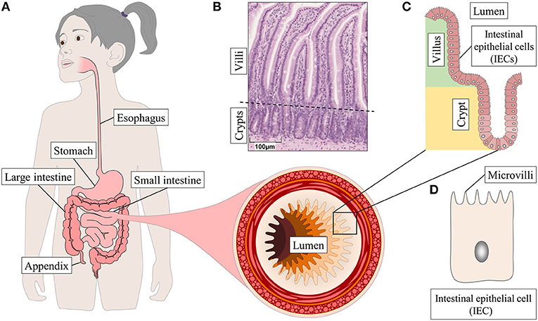
- Figure 1 - (A) The gastrointestinal tract.
- A huge surface area is important for an efficient nutrient absorption. To have the largest possible surface area, the small intestine has big folds that are covered with microscopic fingers” (villi) and “valleys” (crypts). (B) Photo of an intestinal tissue section. (C) Schematic drawing of the crypt-villus structure of the small intestine. (D) The cells that make up the inner surface of the small intestine are called intestinal epithelial cells. These cells also have tiny projections, called microvilli that look like hairs and further increase the surface area.
Box 1 - Experiment - digestion starts in your mouth.
The saliva in your mouth is an example of a digestive juice. You can do a simple experiment to test the enzymes in your saliva. First, you eat a bite of bread (without butter or anything else). Then you thoroughly chew it and keep it in your mouth for a while. It will taste a little bit sweet. Why? Because bread contains starch, which is a carbohydrate. Chemically, carbohydrates like starch are long chains of sugar molecules. They do not taste sweet though, as the sweet taste receptors on your tongue only recognize carbohydrates made of one or two sugar molecules. When the enzymes in your saliva break down the long carbohydrate chains into smaller pieces in your mouth, small sugar molecules are generated that activate the sweet taste receptors, and your tongue tells the brain you ate something sweet.
Digestion breaks down carbohydrates into sugars, proteins into amino acids and fat into tiny droplets called micelles, which contain lipids (Figure 2). IECs take up all these small nutrient components. While micelles can go directly through the cell membrane, sugars, amino acids, and vitamins need special molecules called transporters to enter IECs (Figure 2).
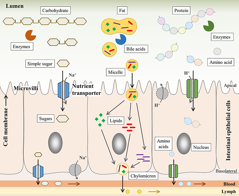
- Figure 2 - The aim of digestion is to break nutrients down into small parts that can be absorbed by intestinal epithelial cells.
- Enzymes break up the chains of sugars and amino acids that build carbohydrates and proteins, respectively. Bile acids act as emulsifiers, like soap, they split fat into tiny droplets, called micelles that can be absorbed directly. Lipids are disassembled in the IECs and re-assembled into chylomicrons. Other nutrients move through the cells using “tunnels,” called transporters. After passing through the IECs, nutrients are distributed throughout the body via the blood and lymph fluid.
What Are Nutrient Transporters?
Transporters form little pores or tunnels in the cell membrane, creating doors through which nutrients can pass. The inner side of the intestine that contains the food mush is called lumen. For absorption, transporters sit on the cell membrane facing the lumen. The reason why nutrients take these doors to leave the lumen and enter the cell is because of diffusion. Diffusion describes a process where molecules move from an area of high concentration (e.g., you ate a muffin and now a lot of sugar is in your intestinal lumen) to an area of low concentration (fewer sugar molecules are in your IECs). The bigger the difference in concentrations, the bigger the physical force that draws the molecules into the cells. Some transporters move ions in parallel to nutrients, using the concentration difference of the ions to fuel nutrient transport. Ions are electrically charged particles, for example salt contains positive sodium and negative chloride ions. But why does the transport of nutrients not slow down or stop? Due to continuous transport, you would expect a decrease in the concentration difference or even the same concentration in the lumen and inside the IECs. To prevent a stop of nutrient transport, IECs also have transporters on their opposite side or “basolateral” side. Using energy, these transporters “pump” out ions, maintaining the low concentration inside the IECs needed for nutrient absorption. Furthermore, nutrient transporters on the basolateral side help nutrients to leave the IECs, so they don’t get overloaded with nutrients. Sugars, amino acids, and vitamins thus pass through the IECs and into the blood stream with the help of transporters. In contrast, fat passes right through the cell membrane, not needing special transporters to do so. Micelles are split into their components in IECs, reassembled into lipid-transport particles called chylomicrons, and are released from IECs into the lymph. All nutrient are then distributed throughout the body and used by all its cells (Figure 2).
Why Do We Need to Study Nutrient Transport?
As you can imagine, nutrient absorption and transport are very complicated processes. Many types of transporters exist that are specific for different types of nutrients, and there are even different transporters for particular types of amino acids and sugars. Also, certain transporters can move more than one type of sugar, and sometimes several types of transporters can move the same nutrient. So, it is no wonder that nutrient absorption is still not fully understood. It is very important to study nutrient transport, because there are diseases that are caused by defective transporters, and some patients have normal transporters but still cannot absorb certain nutrients. For example, patients with fructose-malabsorption, who cannot absorb fruit sugar, often have a functional fructose transporter. Knowing what is wrong in each of these cases could help scientists to find treatments. Treatments are important because malabsorption can cause health problems like severe stomachaches.
Nutrient transporters are also important in drug development, because some medicines are taken up by nutrient transporters. For example, certain antibiotics used to fight bacterial infections are taken up by nutrient transporters, because these drugs have chemical structures similar to small peptides. If we knew the exact details of transport processes, we could invent drugs that can be very efficiently absorbed and cause fewer side effects.
How Is Nutrient Transport Studied?
Unfortunately, it is difficult to study nutrient transport. Scientists study nutrient transport using what are called biological models, because we cannot investigate transport processes directly in the human intestine. Normally, scientists use either cell lines or animals as models, but both have huge drawbacks. Cell lines need to grow and divide constantly so that they will stay alive long enough for the experiment. Therefore, most cell lines originate from tumors or have had their DNA altered. But the changes that make these cells grow so well also change other properties. In particular tumor cell lines, that are very often used to investigate intestinal nutrient transport, sometimes have more transporters than normal cells, or fewer, or they can even lack certain transporters.
On the other hand, in animals you can study more than just absorption of nutrients by IECs. Nutrient uptake can be influenced by factors like the composition of food, the speed with which the food mush passes the villi, and how fast nutrients are passed on to the blood on the basolateral side. This is why it is important to consider the whole organism and not just certain cells. However, if you think of a mouse, the most common animal model—they are rodents, they eat completely different from humans, and their GI tract has a different structure. And even though nutrient transporters of mice and human are similar, they are not the same. Nonetheless, scientists used these models for years, until… a new, groundbreaking method was developed, intestinal organoid culture!
What Are Organoids?
Organoids are small, organ-like structures made of cells. They do not grow as a “cell-lawn” like normal cell lines do, they grow into 3D structures in a substance that resembles jello. Intestinal organoids are like “mini-guts” and they can be grown from crypts (Figure 3) [2]. As the crypts grow in the culture dish, they close and form a balloon, with the IECs forming the wall of the balloon that faces a central lumen, which is like the air inside the balloon. Over time, the organoid balloon starts budding and develops to an octopus-like shape. The numerous arms of the octopus and the areas in between are like the crypts and villi of the small intestine (Figure 3).
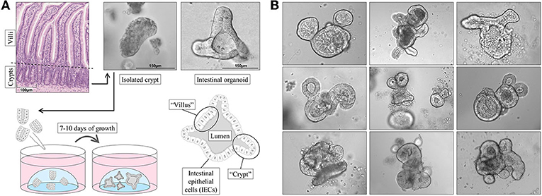
- Figure 3 - (A) Intestinal organoids can be grown from isolated crypts in 7–10 days.
- These “mini-guts” are made of IECs and are a great tool for research. They range in size from the width of a hair (100 μm) up to more than 2 mm, and are visible as small dots even without a microscope. (B) Organoids are also beautiful. Sometimes, if you look through the microscope it seems that they are looking back at you. Organoids can have all kinds of shapes. With a little bit of imagination, you can find a lot in your petri dish! What do you see?
Only very small pieces of intestinal tissue are necessary to get enough crypts to grow organoids. How do we get them? There are two options. Sometimes patients need surgery to have parts of their intestines removed, mainly because of intestinal inflammation or cancer. In these cases, some of the healthy tissue next to the damaged part is removed, too. This tissue can be used to obtain crypts. Biopsies are the other option. A biopsy is a small piece of tissue that can be taken from the GI tract during a medical examination. Biopsies are taken using small clamps that bite into the intestinal wall to get tissue pieces of ~ 10 mm2. To compare, a stud from a LEGO brick has an area of 18 mm2. Out of 2–3 of these tissue pieces, we can obtain 150–300 crypts, which is enough to start growing intestinal organoids [3].
Why Are Organoids Such a Great Model?
Organoids have many advantages:
- they consist of IECs that are exactly like those in the intestine.
- unlike cell lines, they do not originate from cancer or have their DNA altered.
- they are of human origin and not from animals such as mice.
- they can be grown from various parts of the intestine—this is important, because some nutrients are only taken up in certain areas of the intestine, and the necessary transporters are only found in those intestinal areas.
- they can be grown from individual patients and used to find the best treatment for each specific patient. This is called “personalized medicine.”
Conclusion and Outlook
There is still a lot to explore about nutrient absorption in the GI tract. Intestinal organoids are a great tool for this area of research and they will help scientists to better understand diseases like nutrient malabsorption. Using human intestinal organoids as a model system, we now have a better way to test drug absorption through nutrient transporters, which helps with drug development. Last but not least, intestinal organoids will save the lives of laboratory animals, because using human organoids for experiments will reduce the number of experiments performed on animals.
Glossary
Gastrointestinal (GI) Tract: ↑ A long, twisting tube of joined hollow organs: mouth, esophagus, stomach, small intestine, large intestine, and anus. Together with the liver, gall bladder, and pancreas, which produce digestive juices, the GI tract forms the digestive system.
Digestive Juices: ↑ For example saliva, stomach acid, and bile. Chemically (acid and denaturation) and physically (bile and emulsification) break down nutrients and contain enzymes (saliva and pancreatic fluids) to split carbohydrates, proteins, and fat.
Villus/Villi/Microvilli: ↑ Villus: A microscopic, finger-like projection into the lumen made of IECs. Villi: Plural of villus. Microvilli are hair-like structures on the luminal side of IECs, enlarging IEC surface for absorption.
Lymph: ↑ A colorless fluid that is formed when blood fluid (called plasma) exits blood vessels. Fat from the intestines and immune cells travel with the lymph. The main lymph vessel merges with a big blood vessel in the chest, and lymph and blood are blended again.
Biological Model: ↑ Experimental systems that represent tissue functions or diseases in a simplified way. Models respond to medical tests similar to the natural tissue and provide insight into complex biological functions.
Cell Line: ↑ Cells of plants, animals, or humans grown in small plastic containers and used as biological models. They “swim” in medium, a liquid that contains everything the cells need to live and grow.
Personalized Medicine: ↑ The same disease (symptoms) can have different causes. Personalized medicine takes this into account and matches the treatment to each patient individually, to have the best possible treatment.
Conflict of Interest
The authors declare that the research was conducted in the absence of any commercial or financial relationships that could be construed as a potential conflict of interest.
Acknowledgments
ER would like to thank her children for helping her choosing the organoid pictures.
Original Source Article
↑Zietek, T., Giesbertz, P., Ewers, M., Reichart, F., Weinmüller, M., Urbauer, E., et al. 2020. Organoids to study intestinal nutrient transport, drug uptake and metabolism—Update to the human model and expansion of applications. Front. Bioeng. Biotechnol. 5:577656. doi: 10.3389/fbioe.2020.577656
References
[1] ↑ Helander, H. F., and Fandriks, L. 2014. Surface area of the digestive tract—revisited. Scand. J. Gastroenterol. 49:681–9. doi: 10.3109/00365521.2014.898326
[2] ↑ Sato, T., Vries, R. G., Snippert, H. J., van de Wetering, M., Barker, N., Stange, D. E., et al. 2009. Single Lgr5 stem cells build crypt-villus structures in vitro without a mesenchymal niche. Nature 459:262–5. doi: 10.1038/nature07935
[3] ↑ Mahe, M. M., Sundaram, N., Watson, C. L., Shroyer, N. F., and Helmrath, M. A. 2015. Establishment of human epithelial enteroids and colonoids from whole tissue and biopsy. J. Vis. Exp. 97:52483. doi: 10.3791/52483